1Inserm, UMR 1099, Université de Rennes 1, LTSI, Rennes, France, 2Department of Neuroradiology, Pierre-Zobda-Quitman Hospital, University Hospital of Martinique, French West Indies, France, Fort-de-France, France, 3CRLCC, Centre Eugène Marquis, Rennes, France
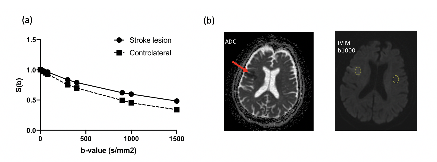
DKI-IVIM analysis on one individual slice of diffusion-weighted images (b = 1000 s/mm ).
(a) Signal intensity plotted as function of b-value for the two ROIs located in the ischemic lesion and normal contralateral region. The slope related to fD* between b = 0s/mm and b = 300 s/mm is decreased for the ischemic region.
(b) Free-hand region of interest (ROI) delineation in the ischemic lesion and the corresponding contralateral ROI on normal tissues.
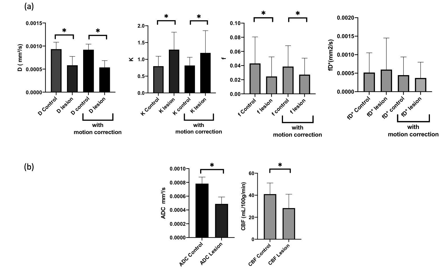
Intravoxel incoherent motion (IVIM) parameters (D, f and fD*) and conventional apparent diffusion coefficient (ADC) and the cerebral blood flow (CBF) mesured through ASL in the lesion and the controlateral normal region averaged over all the stroke patients. Comparison with motion correction data processing for the DKI-IVIM analysis.