Helen Harvey1
1MRI, Cambridge University Hospitals, Cambridge, United Kingdom
1MRI, Cambridge University Hospitals, Cambridge, United Kingdom
This
poster aims to show the techniques for MRI arthrography of the Sternoclavicular
joint (SCJ), review anatomy and demonstrate common pathologies shown with SCJ
arthrography. Helping to increase knowledge and greatly improve treatment and
surgical planning options for the patient.
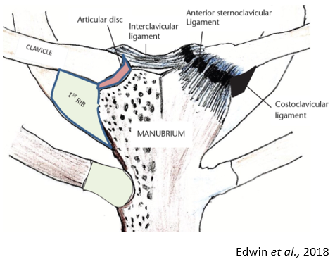
Figure 1: Sternoclavicular joint anatomy