CHIKARA NODA1, Chia Ying Liu2, Jason Ortman1, Bharath Venkatesh Ambale3, Webster Stayman4, Yoshimori Kassai5, and Joao A.C. Lima1
1Cardiology, Johns Hopkins University, Baltimore, MD, United States, 2Canon Medical Research, Cleveland, OH, United States, 3Radiology, Johns Hopkins University, Baltimore, MD, United States, 4Biomedical Engineering, Johns Hopkins University, Baltimore, MD, United States, 5Canon Medical Systems Corporation, Otawara, Japan
1Cardiology, Johns Hopkins University, Baltimore, MD, United States, 2Canon Medical Research, Cleveland, OH, United States, 3Radiology, Johns Hopkins University, Baltimore, MD, United States, 4Biomedical Engineering, Johns Hopkins University, Baltimore, MD, United States, 5Canon Medical Systems Corporation, Otawara, Japan
3D radial Ultrashort echo time sequence is a promising technique for lung imaging but the protocol has not been optimized. We examined the parameters including number of trajectories and flip angles using a lung phantom. In-vivo images were obtained and evaluated based on the parameters optimized in the phantom study. UTE acquisition in axial rather than coronal direction produced better quality of images. Breath holding acquisition may be an option in case patients cannot tolerate long scan time.
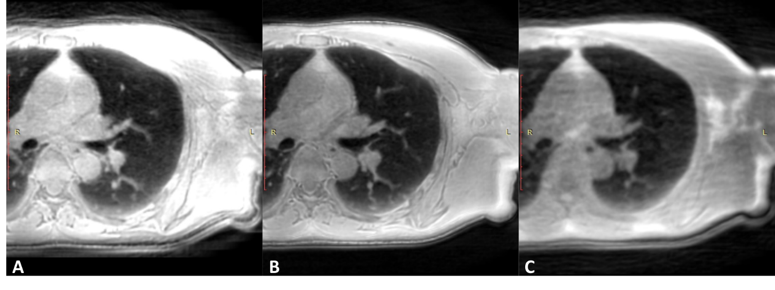
Figure 3. Lung images using ultrashort echo time sequence by different acquisitions. A) post reconstructed to axial by coronal acquisition with respiratory gating. The signal intensity was higher due to the longer acquisition time in this case. B) performed by axial acquisition with respiratory gating. Images were sharper in this orientation. C) performed by axial acquisition with 20 seconds breath holding.