Brian Johnson1, Jonathan Chia1, Dave Hitt1, Rob Lay1, Tom Lowe1, Michael Pawlak1, John Penatzer1, James Snicer1, Marcie Stopchinski1, Gregory Thomas1, Kristen Williams1, and Paul Worthington1
1Philips Healthcare, Gainesville, FL, United States
1Philips Healthcare, Gainesville, FL, United States
Here we
present an optimized radial k-space sampling protocol of the cervical spine.
Use of radial k-space filling techniques helps reduces motion artifacts and
leads to a more comprehensive scanning strategy to provide consistent and high image
quality.
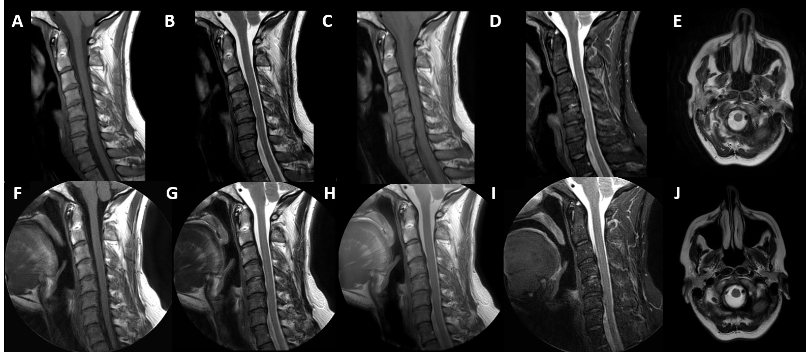
Figure 3: Examples of 2D cartesian cervical spine image contrasts
outlined in the ACR guidelines (A) sagittal T1-weighted (B) sagittal
T2-weighted (C) sagittal proton density (D) sagittal STIR and (E) axial
T2-weighted compared to radial acquisitions for (F) sagittal T1-weighted (G)
sagittal T2-weighted (H) sagittal STIR (I) Axial T2-weighted, and axial (J)
T2-weighted images.