Kevin Epperson1, Karla Epperson1, Garry Gold2, and Feliks Kogan3
1Radiology School of Medicine, Stanford University, Stanford, CA, United States, 2Radiology, Stanford Univeristy, Stanford, CA, United States, 3Radiology - SOM, Stanford Univeristy, Stanford, CA, United States
1Radiology School of Medicine, Stanford University, Stanford, CA, United States, 2Radiology, Stanford Univeristy, Stanford, CA, United States, 3Radiology - SOM, Stanford Univeristy, Stanford, CA, United States
Chronic knee pain, invading osteoarthritis, and rising diagnostic costs continue to hinder diagnosis and treatment of this disease. Can the benefits of high field MR imaging such as 7T provide the needed resolution to present pathology as well as allow the quantitative results expected.
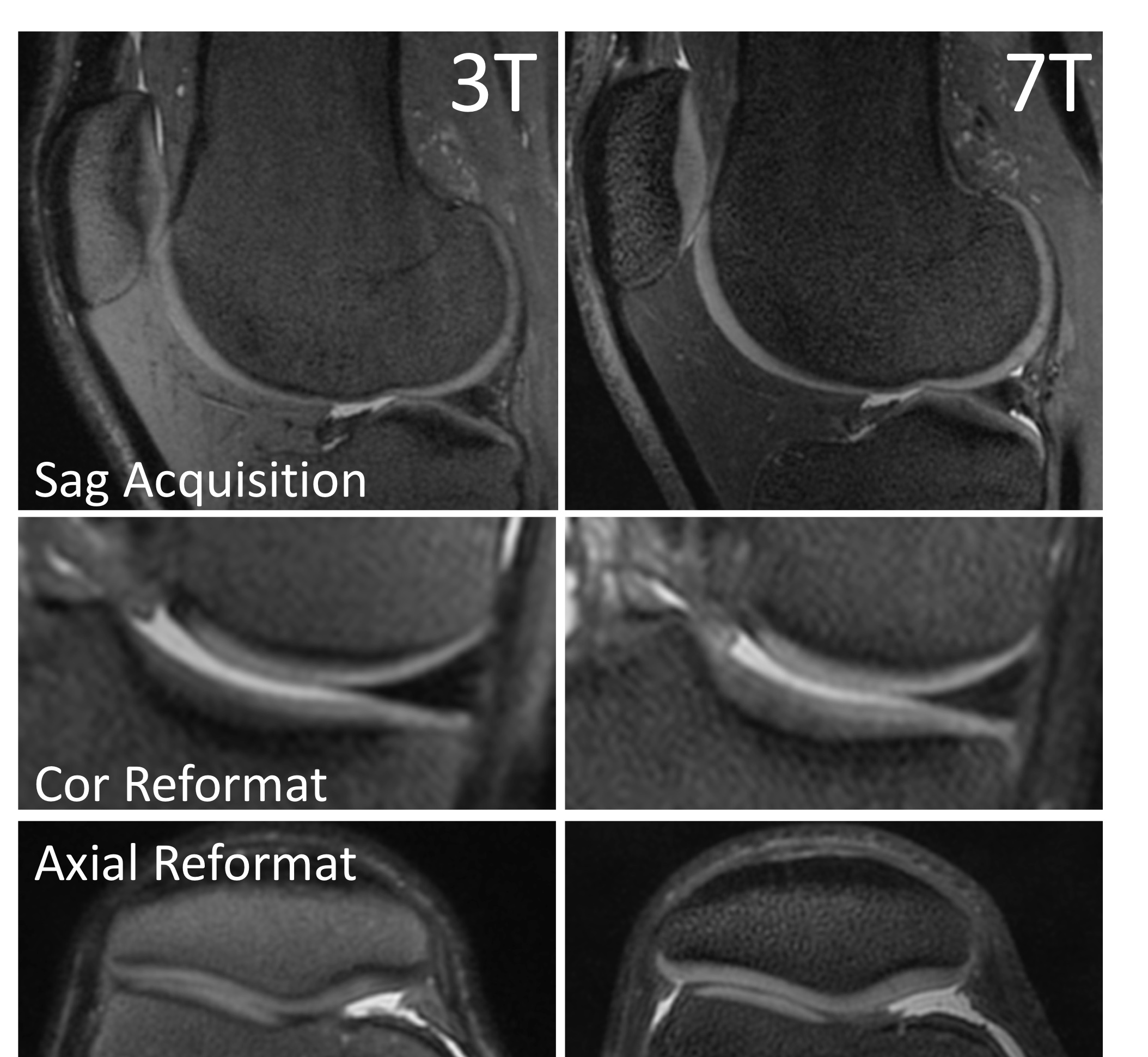
Figure 2. 3D PD Cones images acquired at 3T (left) and 7T (right) with equivalent parameters. 7T images shown considerably better tissue signal and contrast in the deep layers of patellar and tibial cartilage. Additionally, on coronal and axial reformats, 7T images show improved visualization of structures with reduced blurring.