Weekday Course
Standing Out: Contrast Mechanisms in Breast Imaging
ISMRM & SMRT Annual Meeting • 15-20 May 2021

Weekday Course
Standing Out: Contrast Mechanisms in Breast Imaging
| Concurrent 7 | 15:00 - 16:00 | Moderators: Hai-Ling Cheng & C. C. Tchoyoson Lim |
 |
Perfusion Imaging of the Breast
Wei Huang
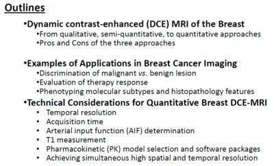
Perfusion imaging of the breast as measured by the most common method of DCE-MRI is reviewed in this lecture. The three approaches for breast DCE-MRI data analysis, qualitative curve shape description, semi-quantitative analysis, and quantitative pharmacokinetic modeling, as well as their pros and cons, are discussed. Examples of major applications in clinical care and research of breast cancer are demonstrated. With quantitative pharmacokinetic modeling of DCE-MRI data increasingly used in clinical trial and research settings, major technical considerations that affect quantitative parameter accuracy and precision are discussed.
|
|
 |
Spectroscopy in Breast Imaging
Uma Sharma
In-vivo proton proton MR spectroscopy (MRS) has emerged as a non-invasive tool for diagnosis and to provide an insight into the biochemistry of breast cancer. The elevated levels of choline containing compounds (tCho) have been identified as a non-invasive biomarker for differentiating malignant and benign breast lesions. Using in-vivo quantification of absolute tCho concentration, cut-off values for the differentiation of malignant, benign and normal breast tissues were estimated. Combined use of water-to-fat ratio (W-F), tCho and lipid resonances improved the diagnostic ability of breast MRS. Monitoring of tCho and W-F ratio has been useful in predicting therapeutic response of tumor.
|
|
 |
HIFU & Breast Interventions
L. Wilbert Bartels
Bioeffects provoked by ultrasound can be exploited for therapeutic use. For interventions in the breast, the thermal effects of high intensity focused ultrasound are of particular interest, both for thermal ablation of lesions, as well as for delivering long term mild hyperthermia treatments. MRI is well suited for image guidance of HIFU therapy, as it combines imaging with excellent soft-tissue contrasts with the ability to map and monitor temperature changes during therapy. Experiences with an MR-HIFU system specifically designed for breast applications will be discussed, with particular attention for challenges related to MRI for therapy planning, guidance, and evaluation.
|
|
 |
Diffusion-Weighted Imaging of the Breast
Savannah Partridge
Diffusion-weighted imaging (DWI) holds promise to address shortcomings of routine clinical breast MRI and to expand imaging capabilities in breast cancer management. DWI reflects tissue microstructure and provides unique information to aid in detection and characterization of breast lesions. Potential applications include improving diagnostic accuracy, guiding treatment decisions, and non-contrast screening. DWI is increasingly being incorporated into breast MRI protocols, and results of multicenter trials and recent standardization efforts are helping to establish clinical guidelines. Advancements in DWI acquisition and modeling approaches are emerging to improve image quality and extract additional biologic information from breast DWI scans.
|
The International Society for Magnetic Resonance in Medicine is accredited by the Accreditation Council for Continuing Medical Education to provide continuing medical education for physicians.
 Watch the Video