Weekend Course
MRI in Sports Imaging: Lower Extremity
ISMRM & SMRT Annual Meeting • 15-20 May 2021

Weekend Course
MRI in Sports Imaging: Lower Extremity
| Concurrent 2 | 13:00 - 13:45 | Moderators: Section 1: Shivani Ahlawat & Richard Kijowski Section 2: Garry Gold & Hiroshi Yoshioka |
| Section 1 | ||
 |
MRI of the Muscle-Tendon Unit
Joon-Yong Jung
The muscle-tendon unit is an axis to transmit force from muscle to skeleton, and its structural integrity is a prerequisite to preserve locomotion. Myotendinous injuries frequently occur not only in elite athletes, but also in the general population participating in recreational activity. This presentation reviews the microscopic and macroscopic anatomy of muscle-tendon unit in correlation with MR findings, the diagnostic approach to myotendinous injury in lower extremities using MRI, and imaging parameters associated with longer return to play or reinjury. Additionally, promising MR techniques for assessment of myotendinous injury will be briefly touched upon.
|
|
 |
MRI of the Athletic Hip
Adam Johnson
MRI plays a vital role in the management of hip pain in active patients. Using several cases, we will illustrate the MRI findings in some common and less common causes of hip pain. We will dive a bit deeper into the various forms of hip impingement including both intra and extra-articular causes and their associated pathology. When appropriate, we will discuss the benefit of MR arthrography over standard MRI. We will also review the recommendations from the Society of Skeletal Radiology (SSR) Subchondral Bone Nomenclature Committee as they pertain to the hip.
|
|
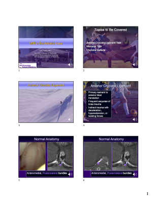 |
MRI of the Athletic Knee
Erin Alaia
In this lecture, the viewer will learn about common pathology seen on MRI of the athletic knee. The lecture will cover the MRI appearance of anterior cruciate ligament tears, meniscal tears, and knee chondral defects, as well as commonly observed associated and ancillary imaging abnormalities. Basic anatomic and functional considerations will be highlighted, along with arthroscopic correlation, in order for the viewer to better understand the MRI imaging appearance and clinical implications.
|
|
| Section 2 | ||
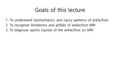 |
MR imaging of Sports injuries of the Ankle & Foot
Jung-Ah Choi
In this lecture, we will learn about the biomechanics and injury patterns of the ankle and foot in sports injuries. We will learn how to diagnose common sports injuries of the ankle and foot on MRI, including medial and lateral, Lisfranc ligamentous injuries, Achilles tendon tears, stress fractures, and osteochondral lesions, with recognition of limitations and pitfalls of MRI.
|
|
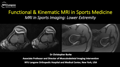 |
Functional & Kinematic MRI in Sports Medicine
Christopher Burke
Pain due to patellofemoral maltracking and femoroacetabular impingement are common reasons for presentation to sports medicine clinics. These dynamic phenomena also both represent independent risk factors for early onset osteoarthritis. Patients are usually evaluated with clinical examination supported by radiographs and standard MRI with joint held in a static position. In addition to standard MRI protocols, additional kinematic sequences can be used in everyday orthopedic practice to aid the clinical work up and assessment of these patients. The background and potential application of these techniques are presented.
|
|
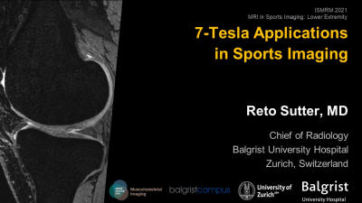 |
7-Tesla Applications in Sports Imaging
Reto Sutter
7T imaging can be a powerful tool for sports imaging. While for many athletes it is possible to obtain a correct diagnosis at lower field strengths, some athletes may benefit from higher spatial resolution, and it is possible to identify use cases for sports imaging at 7T. MRI at ultra-high field strengths comes with technical challenges such as field homogeneity and fat/water separation, but these can be successfully overcome to enable high resolution imaging. Most work has been done for knee imaging and muscle imaging, but pilot studies for hip and foot imaging at 7T are available.
|
|
The International Society for Magnetic Resonance in Medicine is accredited by the Accreditation Council for Continuing Medical Education to provide continuing medical education for physicians.
 Watch the Video