Weekend Course
MRI in Sports Imaging: Upper Extremity
ISMRM & SMRT Annual Meeting • 15-20 May 2021

Weekend Course
MRI in Sports Imaging: Upper Extremity
| Concurrent 2 | 13:45 - 14:30 | Moderators: Section 1: Jan Fritz & Hollis Potter Section 2: Kimberly Amrami & Robert Boutin |
| Section 2 | ||
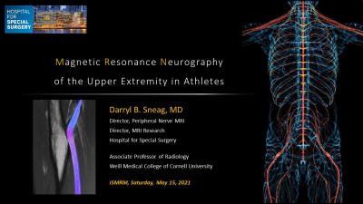 |
MR Neurography of the Upper Extremity in Athletes
Darryl Sneag
This presentation reviews general concepts related to acquisition and interpretation of magnetic resonance (MR) neurography exams. MR neurography is an important adjunct to both the physical exam and electrodiagnostic testing in the evaluation of peripheral nerve injuries in athletes. MR neurography is non-invasive and provides higher contrast resolution and access to deeper nerves/muscles, as compared to ultrasound, which is also highly operator-dependent. MR neurography of the upper extremity encompasses injuries extending from the brachial plexus to the fingers, but most athletic injuries in clinical practice that are referred for imaging are centered around the neck and shoulder regions.
|
|
| Sports Injuries in Adolescents
Jie Nguyen
Increasing participation in youth sports has led to the growing incidence of acute and overuse injuries. In skeletally immature children, growth plate is the weak link, producing a spectrum of findings ranging from reversible physeal widening to irreversible adaptive remodeling, physeal fracture, and premature physeal closure. If undiagnosed and untreated, these changes can lead to deformity and premature osteoarthritis. This presentation will review the normal growth plate complex and highlight physeal-specific pathologies that can occur in the shoulders and knees of youth athletes (proximal humeral epiphysiolysis, physeal and avulsion fractures, glenoid remodeling, osteochondritis dissecans, and transphyseal bar). |
||
| Section 1 | ||
| Biomechanics of Sports Injuries
James Johnston
|
||
| MRI of the Athletic Elbow, Wrist & Hand Video Permission Withheld
Amanda Isaac
|
||
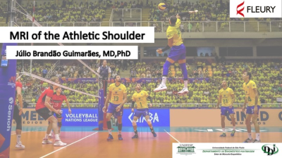 |
MRI of the Athletic Shoulder
Júlio Guimarães
Shoulder disease is common in the athletic population. The unparalleled velocity achieved by overhead throwers subjects the shoulder to extreme forces, resulting in adaptive changes and pathologic findings that can be detected at imaging.A key biomechanical principle of throwing is achieving maximum external rotation, which initially leads to adaptive changes that may result in a pathologic cascade of injuries. Magnetic resonance imaging most important imaging modality to athletic shoulder pathologies and knowledge of injury patterns specific to throwing shoulder help radiologist identify the total spectrum of abnormalities and provide more relevant clinical insight to treating orthopedic surgeon.
|
|
| Section 2 | ||
| MRI of Treatment & Recovery
C. Benjamin Ma
|
||
The International Society for Magnetic Resonance in Medicine is accredited by the Accreditation Council for Continuing Medical Education to provide continuing medical education for physicians.
 Watch the Video