Weekend Course
Cardiovascular MRI: From Disease to Diagnosis
ISMRM & SMRT Annual Meeting • 15-20 May 2021

| Concurrent 1 | 13:45 - 14:30 | Moderators: Daniel Kim & Maria Drangova |
| Cardiovascular Disease: Macro- & Micro-Vasculature
Christopher Francois
|
||
| Myocardial Disease: Pathophysiology & Unmet Diagnostic Needs
Andrew Arai
|
||
 |
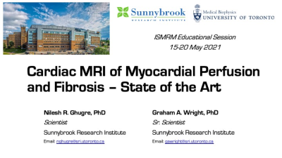
Cardiac MRI of Myocardial Perfusion & Fibrosis - State of the Art
Graham Wright, Nilesh Ghugre
Assessment of perfusion and fibrosis in the heart with MRI is central to a complete cardiac exam. The core clinical methods have focused on dynamic first pass of Gadolinium-based contrast for perfusion and late gadolinium enhancement for fibrosis. Newer methods have targeted improvements in spatial resolution and coverage with reductions in imaging time, and robustness in the presence of implanted devices. There is a growing emphasis on quantitatiion and the introduction of methods that do not require the injection of a contrast agent. Clinical applications include management of ischemic and non-ischemic heart disease as well as complex arrhythmia management.
|
|
 |
Measuring Ventricular Microstructure & Strain
Daniel Ennis
Cardiac structure and function are inextricably bound. MRI is an unrivaled technology for exploring and discovering the structure-function mechanisms of cardiac function and dysfunction. This talk reviews the basic microstructural constituents of the hearts (i.e. “myofibers” and “sheets”), two methods for measuring regional cardiac function (i.e. MRI tagging and cine DENSE), and the use of cardiac diffusion tensor MRI to measure microstructural organization in vivo. Methods to jointly integrate measures of structure and function to reveal microstructurally anchored measures of cardiac function (.e.g “myofiber” strain) are also described.
|
|
 |
Tissue Characterization: Relaxometry & Exchange
Weitian Chen
Relaxation time constant T1, T2, and T1rho are important parameters to characterize tissue properties. The spatial and temporary variation of the magnetic field due to dipolar coupling and molecular tumbling is a main source of the observed relaxation effect. Relaxation can also be affected by chemical exchange and magnetization transfer effect. These physical processes reflect the metabolites and macromolecules content in tissues. Thus, imaging methods based on the measurements of relaxation time can be used to probe the biochemical properties of tissues.
|
|
| Outcomes of Trials for Quantitative CMR Biomarkers
Michael Salerno
|
The International Society for Magnetic Resonance in Medicine is accredited by the Accreditation Council for Continuing Medical Education to provide continuing medical education for physicians.