Weekend Course
Vascular Pathology & Imaging
ISMRM & SMRT Annual Meeting • 15-20 May 2021

| Concurrent 1 | 14:30 - 15:15 | Moderators: Toshimasa Clark & Chengcheng Zhu |
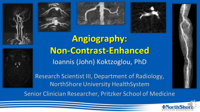 |
Angiography: Non-Contrast-Enhanced
Ioannis Koktzoglou
This presentation overviews non-contrast-enhanced techniques for magnetic resonance angiography (MRA). Non-contrast-enhanced MRA can be performed using a variety of techniques. This presentation describes the main non-contrast-enhanced MRA techniques, their contrast mechanisms, as well as their clinical applications. Strengths and drawbacks of non-contrast-enhanced MRA will also be discussed. This presentation also reviews emerging methods that hold promise for expanding the role of non-contrast-enhanced MRA in the clinical setting.
|
|
 |
Molecular targeting of inflammation in atherosclerotic vascular disease
Katey Rayner
Atherosclerosis is the disease that causes heart attack and stroke. Although driven by excess cholesterol, atherosclerosis is a chronic inflammatory disease. Macrophages engage inflammatory pathways such as the inflammasome and activate NFkappa B transcriptional of cytokines to propagate inflammation, and new clinical data suggests that targeting these pathways therapeutically holds promise for patients with CVD. We will discuss new advances in inflammatory pathways in the advanced atherosclerotic plaque and how they may be leveraged for imaging of advanced atherosclerosis.
|
|
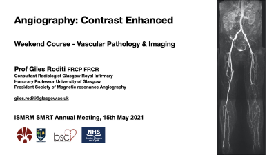 |
Angiography: Contrast-Enhanced
Giles Roditi
This presentation focuses on the basic principles of contrast-enhanced magnetic resonance angiography with emphasis on these foundations: 1 - Administration of intravenous contrast agent to provide contrast by shortening T1 of blood in the vessel lumen relative to background tissues 2 - Timing of the scan to coincide with contrast agent arrival in vascular bed of interest using bolus tracking or test bolus methods 3 - Scan parameters that influence image quality CE-MRA can be performed in first pass, dynamically, in ’extended phase’ (equilibrium / steady state) or combinations thereof, the advantages and trade-offs involved in each will be explored. |
|
 |
Markers of Vascular Function
Jos Westenberg
Vascular function testing may provide insight in early subclinical disease state. Understanding blood flow characteristics near the wall is relevant when testing vascular function for diagnosing and predicting future risk of cardiovascular disease. Phase Contrast MRI assesses blood flow velocity and provides unprecedented hemodynamic insight when extended to 4D Flow MRI. Relevant markers of vascular function assessed by 4D Flow MRI are: vascular wall stiffness (Pulse Wave Velocity), hemodynamic force on the wall (Wall Shear Stress) and eccentricity in blood flow pattern (Flow displacement).
|
|
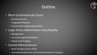 |
Vessel Wall Imaging: Extracranial
Mahmud Mossa-Basha
Extracranial vasculopathy assessment, has primarily focused on carotid atherosclerotic disease, as this represents the most common extracranial vasculopathy. In the current lecture, we evaluate less frequently discussed extracranial vasculopathies, specifically blunt cerebral vascular injury and dissection and large artery inflammatory vasculopathies. We discuss disease background, current limitations in imaging paradigms and the value of vessel wall MRI, represented through case examples. In addition, we briefly discuss carotid atherosclerotic disease, providing examples of pronounced plaque burden but with limited luminal stenosis, and discuss automated, quantitative algorithms for plaque evaluation, that may improve clinical adoption of vessel wall MRI in plaque characterization.
|
|
 |
Vessel Wall Imaging: Coronaries
Reza Hajhosseiny
Coronary artery disease (CAD) remains a significant cause of mortality and morbidity worldwide. Conventional population derived risk stratification tools, coronary luminography and functional assessment of CAD all have significant limitations in identifying individual patients most at risk of major adverse cardiac events. Coronary vessel wall imaging is a promising tool for the timely detection of coronary atherosclerosis. Advantages include early individualised risk stratification, bespoke and targeted therapeutical intervention and monitoring the response to treatment and disease progression. Cardiovascular magnetic resonance, computed tomography and nuclear imaging are all at the forefront of non-invasive coronary vessel wall and vulnerable plaque imaging.
|
|
| Vessel Wall Imaging: Intracranial
Jae Song
This lecture discusses intracranial vessel wall MR imaging and how to work towards translating evolving and innovative technologies into the clinical sphere. We highlight that it is important not only to design new technologies and showcase what the MR community can innovate but also to translate that technology into a stage of implementation and adoption. We discuss technical unmet needs and potential clinical applications. We review the importance of engagement and collaboration with clinicians to ensure longevity of the research by prioritizing relevant research questions in the context of feasibility.
|
The International Society for Magnetic Resonance in Medicine is accredited by the Accreditation Council for Continuing Medical Education to provide continuing medical education for physicians.
 Watch the Video