Weekend Course
Neurodevelopmental Disorders
ISMRM & SMRT Annual Meeting • 15-20 May 2021

| Concurrent 2 | 14:30 - 15:15 | Moderators: Mai-Lan Ho |
 |
Neurodevelopmental Disorders: Genetics & Neurophysiology
Mai-Lan Ho
This talk will cover MRI of brain development, including age-related neurodevelopment, normal variants, and diagnostic pitfalls, and key findings in neurogenetic disorders. We will review key mechanisms of pediatric brain development based on major embryologic steps including dorsal induction, ventral induction, formation of midline commissures, migration, and organization. We will cover MRI of fetal, perinatal, and postnatal development, including myelination, sulcation, basal ganglia, pituitary, and ventricular findings. Multiple imaging examples of congenital brain malformations will be presented, linking MRI findings to underlying derangements of neurodevelopmental processes.
|
|
 |
Autism & Sensory Over-Responsivity: Linking Structural & Functional Abnormalities
Susan Bookheimer
_
|
|
| Commissures of the Brain Video Permission Withheld
Ajay Taranath
Bundles of white matter that connect homologous structures of the cerebral hemispheres are designated commissures. The hippocampal commissure anterior commissure and fornix are the other commissures. The corpus callosum has evolved as a response to the demand for a shorter connecting route between the rapidly expanding cerebral hemispheres in placental mammals. It is the commissure of the neocortex.
|
||
 |
Brainstem & Cerebellar Malformations
Kshitij Mankad
In this brief presentation, we review how both embryological and environmental factors can create a diverse spectrum of malformations involving the posterior fossa of the brain. We first review entities where the neuroimaging phenotyping is strong and well evolved, matching these to the clinical phenotype as well as genetic bases (where known); We then look at some novel entities with distinct phenotypes, as well as new mechanistic insights and philosophies into existing conditions. Associated supratentorial malformations are discussed and described where relevant. In addition, we review the role of diffusion tensor imaging (DTI) in entities where the technique adds value.
|
|
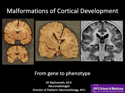 |
Cortical Malformations: From Gene to Phenotype. Video Permission Withheld
Alireza Radmanesh
Understanding cortical development and embryology is essential to have a deep understanding of cortical malformations. Cortical malformations are classically categorized based on the stage of cortical development that is impacted: Proliferation, migration, late migration/organization. With our increasing understanding of molecular pathways and genetics, it has come to light that many different genes can lead to a similar MRI appearance (phenotype) because they are parts of the same molecular pathway. Therefore, understanding molecular pathways is more helpful than memorizing genes in attempts to understand disease conditions.
|
|
 |
Imaging Brain In Utero, Anatomical & Functional: Techniques, Problems & Solutions
Esra Abaci Turk
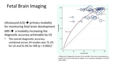
Although ultrasound is still the primary modality for monitoring developing brain in utero, MRI has become available for pregnant patients as a secondary screening tool increasing the diagnostic accuracy achievable by ultrasound. Clinical MRI protocols usually relies on fast acquisition techniques. Two main challenges in fetal MRI are motion artifacts and safety concerns. Even though there are novel motion correction approaches, this topic continues to be a growing research area. There is no report on harmful effect of MRI to the growing fetus. However, fetal MRI safety at each gestational age, particularly the first trimester, is under investigation.
|
|
| Neurophakomatoses
Matthew Whitehead
Neurophakomatoses comprise a diverse group of disorders affecting structures derived from ectoderm and/or mesoderm, leading to skin and intracranial abnormalities. MRI plays an important role in diagnosis, prognosis, and surveillance. Frequently, the imaging pattern is diagnostic or highly suggestive; the radiologist may be the first to suggest the diagnosis. In this lecture, I will attempt to strike a balance between depth and breadth, beginning with detailed examples of the 5 most common phakomatoses including neurofibromatosis type 1, Tuberous sclerosis, Sturge-Weber syndrome, Von-Hippel Lindau, and neurofibromatosis type 2, with a rapid review of rarer neurophakomatoses with specific neuroimaging patterns to follow.
|
The International Society for Magnetic Resonance in Medicine is accredited by the Accreditation Council for Continuing Medical Education to provide continuing medical education for physicians.
 Watch the Video