Weekend Course
Relaxation: Principles & Measurement Techniques
ISMRM & SMRT Annual Meeting • 15-20 May 2021

Weekend Course
Relaxation: Principles & Measurement Techniques
| Concurrent 4 | 15:15 - 16:00 | Moderators: Yun Jiang & Teresa Nolte |
| Physical & Physiological Principles of Relaxation
Sean Deoni
|
||
 |
Methods for T1 & T1rho Mapping
Nikola Stikov
With advances in hardware and the growing availability of post-processing tools, it is becoming easier and faster to obtain accurate T1 and T1ρ maps in clinically feasible times. This course shines a light on the three basic types of T1 mapping techniques, exemplified by inversion recovery, variable flip angle, and MP2RAGE. The course will also introduce techniques for quantitative mapping of the spin-locked relaxation time (T1ρ). T1ρ is closely related to both T1 and T2, but sensitive to different properties of the tissue, and has therefore garnered interest in specialized applications (e.g., cartilage imaging).
Interactive tutorial available at: http://qmrlab.org/t1_book |
|
 |
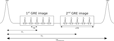
Methods for T2 & T2* Mapping
Richard Dortch
Researchers have developed an array of methods for mapping T2 and T2* values in tissue. The choice of the method depends on numerous factors, including the signal model (single versus multi-compartment), scan time, SNR, and the tissue of interest. In all cases, care must be taken to optimize sequence parameters and minimize the impact of confounding features (B0 and B1 variations). Moving forward, it is likely that more efficient and robust methods for estimating T2 and T2* in tissue will continue to be developed, especially as multi-compartment models continue to illustrate their ability to quantify microstructural features (e.g., myelin content).
|
|
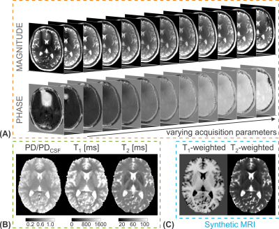 |
Methods for Multiparameter Mapping
Rahel Heule
This talk gives a technical overview about acquisition strategies suited to map longitudinal and transverse relaxation times simultaneously. Special focus is on fast joint T1 and T2 quantification based on three classes: multi-contrast steady-state free precession (SSFP) imaging, magnetization-prepared (MP) schemes with SSFP readout, and magnetic resonance fingerprinting (MRF) acquisitions. Possible acquisition strategies to enhance T2* sensitivity for simultaneous quantification of T1, T2, and T2* are introduced briefly as well.
|
|
| Advanced Reconstruction Methods for Relaxation Parameter Mapping
Alessandro Sbrizzi
Classic relaxation parameter mapping sequences such as inversion-recovery (for T1) or multiple-echo spin-echo (for T2) are too long for clinical applications. By better exploiting structure and relationships (priors) in the spatial (or frequency) domain and in the sequence parameter domain it is possible to under-sample the acquisition thereby accelerating the scan times. More advanced modelling strategies (e.g. time-domain) leads to further acceleration. However, the reconstruction algorithms gets more complex and computationally demanding. Deep learning strategies could overcome these drawbacks.
|
||
 |
Dedicated Cardiac Parameter Mapping Methods
René Botnar
Despite the current success of myocardial tissue characterization with parametric mapping approaches, the accuracy and precision of T1, T2, T2* and ECV mapping can be affected by many confounding factors such as B0 and B1 inhomogeneity, respiratory and cardiac motion, heart rate variability, magnetisation transfer and other parameters. Moreover, estimation of T1, T2 and T2* is usually done by pixel-wise fitting of the MR signal to simple exponential models that may be an oversimplification of the true MR signal evolution. Here we will review the basic myocardial mapping techniques, discuss their pros and cons and subsequently discuss potential solutions.
|
The International Society for Magnetic Resonance in Medicine is accredited by the Accreditation Council for Continuing Medical Education to provide continuing medical education for physicians.
 Watch the Video