Weekend Course
Standardized & Quantitative Assessment in Body Imaging
ISMRM & SMRT Annual Meeting • 15-20 May 2021

Weekend Course
Standardized & Quantitative Assessment in Body Imaging
| Concurrent 1 | 13:00 - 13:45 | Moderators: : Mustafa Shadi Bashir : Silvia Chang & Evis Sala |
 |
Liver: Iron Quantification Video Permission Withheld
Suraj Serai
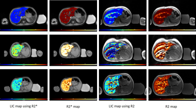
Serial surveillance of liver iron concentration (LIC) provides guidance for chelation therapy in patients with iron overload. The diagnosis of iron overload traditionally relies on core liver biopsy, which is limited by invasiveness, sampling error, cost and general poor acceptance by patients and their family. Thus noninvasive diagnostic methods such as MRI are highly attractive for quantification of liver iron concentration (LIC). In this talk, we discuss and review the MRI based quantification methods of LIC.
|
|
| Liver: Fibrosis Quantification
Verena Obmann
Plenty of etiologies of diffuse liver disease eventually result in liver fibrosis. Liver fibrosis is exceedingly common worldwide prompting the clinical need for objective noninvasive methods for disease characterization as an alternative or complement to gold-standard liver biopsy. The learning objectives of this educational session are • To summarize qualitative imaging findings of liver fibrosis in US, CT and MRI • To know the advantages and drawbacks of each modality • To learn how to report the presence of liver fibrosis
|
||
| Liver: Fat Quantification
S. Sendhil Velan
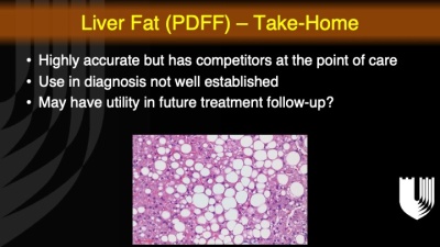
This presentation will cover the MRI/MRS-based techniques for the quantification of liver fat. Specifically, this presentation will include relevant MRI/MRS techniques, types of pulse sequences utilized in a clinical setting, challenges, and finally, the state of the art of development and validation of MRI-based approaches for quantification of liver fat.
|
||
 |
Case Discussion: Liver
Mustafa Shadi Bashir
This talk will discuss the use of MRI-based quantitative biomarkers in the care of patients with liver diseases.
|
|
 |
Bladder: VI-RADS
Valeria Panebianco
The purpose of the talk is to describe the newly developed VI-RADS scoring system aimed at standardization of MRI acquisition, interpretation, and reporting for urinary bladder cancer (BCa). An insight will be given on the BCa diagnostic issues, MRI applications for BCa local staging with assessment of muscle invasiveness, and clinical implications of the score for the disease management. In addition, future prospective of the score applicability will be provided, on its role in patients’ stratification for therapeutic planning, disease surveillance, and for the evaluation of response to therapy. Finally, a clinical series with multiple-choice questions will be shown.
|
|
| Case Discussion: Genitourinary
Caroline Reinhold
|
||
| Kidney: Bosniak
Matthew Davenport
Bosniak v.2019 is a classification of cystic renal masses that formally incorporates MRI, provides verbose specific definitions for imaging terms, and expands the number of masses to which the historic Bosniak classification applies. The intent of the classification update was to improve specificity for malignancy and improve inter-rater agreement, with the goal of reducing the harms of overdiagnosis and overtreatment.
|
||
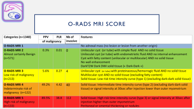 |
Ovary: O-RADS
Isabelle Thomassin-Naggara
This lecture will present O-RADS MR score which was developed to predict malignancy of adnexal masses. This score is an accurate five level categories score that demonstrates a sensitivity and specificity higher than 90% and a good reproducibility (Kappa >0.8). In a multicentric european large validation cohort, only 2 borderline and no invasive cancer were quoted O-RADS MR score 2 (NPV = 98%). This score is mainly based on the analysis of solid tissue using morphological and functional criteria including T2W signal, DW signal and the analysis of time intensity curve on DCE MR sequence.
|
|
The International Society for Magnetic Resonance in Medicine is accredited by the Accreditation Council for Continuing Medical Education to provide continuing medical education for physicians.
 Watch the Video