Weekend Course
Perfusion MRI
ISMRM & SMRT Annual Meeting • 15-20 May 2021

| Concurrent 2 | 14:30 - 15:15 | Moderators: Jongho Lee & Toshinori Hirai |
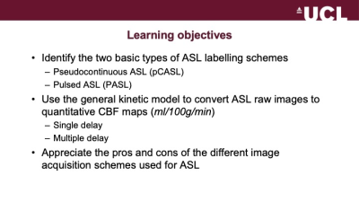 |
ASL: Acquisition & Analysis in the Brain
David Thomas
ASL is a non-invasive MRI method for quantitative mapping of cerebral blood flow (CBF). In this talk, I describe the two main categories of ASL – pseudocontinuous (pCASL) and pulsed (PASL) – and explain how the images acquired using these different acquisition schemes can be converted into CBF maps, using a general tracer kinetic model. I describe how ASL data can be acquired with either single or multiple inflow delay times, and the pros/cons of these two approaches. Lastly, I discuss some practical considerations (imaging method, background suppression) which also affect the quality and accuracy of ASL CBF maps.
|
|
| ASL: Acquisition & Analysis in the Body
Susan Francis
This lecture will outline Arterial Spin Labelling acquisition and analysis methods used in body applications. The considerations for ASL acquisition in terms of labelling schemes and post-label delay, image readout, and methods to reduce motion effects for body ASL are discussed. Analysis methods used in the body applications to account for transit delays and correct for motion will be discussed. The application of ASL MRI across various body organs will be outlined including the kidney, placenta, pancreas and liver, and heart, with clinical applications highlighted.
|
||
 |
ASL: Clinical Applications of ASL
Jeff Winter
Arterial spin labeling (ASL) is emerging as a valuable tool for various clinical applications primarily in the brain, but also in the body. In this session, we will review key clinical applications, including stroke, cerebrovascular disease, neurodegeneration, neuro-oncology as well as renal applications. We will highlight how both cerebral blood flow as well as transit time effects can be used in different clinical applications. Lastly, we will highlight challenges and recent initiatives to increase adoption of ASL perfusion imaging in the clinic.
|
|
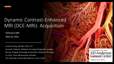 |
DCE-MRI: Acquisition
Caroline Chung
This educational session will review the workflow components of Dynamic Contrast-Enhanced MRI acquisition and will provide an overview of the recommendations and discussions included in the DCE-MRI Quantification Profile of the Quantitative Imaging Biomarker Alliance of the Radiological Society of North America, which has recently wrapped up its public comment phase. This version 2.0 profile update specifically aims to address issues and challenges to consider with 3T MRI and parallel imaging.
|
|
| DCE-MRI: Analysis
Lucy Kershaw
Analysing DCE-MRI data can be time-consuming and complex. In this session, the process will be broken down into steps whilst highlighting potential pitfalls. Analysis and acquisition are closely linked but we will start from the position of having acquired anatomical images, T1 maps and the dynamic series, and assume that analysis will be done using python, matlab etc. We will then cover:
|
||
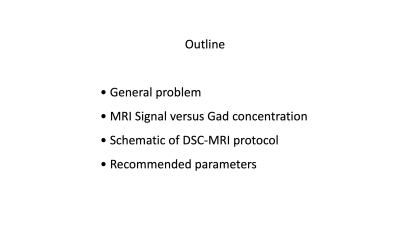 |
DSC-MRI: Acquisition
Greg Cron
This short talk focuses on the bare-bones basics of DSC-MRI acquisition. We will look at the relationship between MRI signal and Gadolinium concentration in a qualitative manner. We will explain how preloading suppresses T1 weighting. We will then show a schematic of a basic DSC-MRI acquisition protocol and provide recommended parameters.
|
|
| DSC-MRI: Analysis
Linda Knutsson
Perfusion is the term applied to capillary blood flow in tissue. The study of brain perfusion has clinical applications due to the changes in perfusion associated with several neurological diseases. Dynamic susceptibility contrast (DSC) MRI is a method for retrieving perfusion and perfusion-related parameters using an exogenous contrast agent. The quantification of the perfusion and perfusion-related parameters from DSC-MRI is a two-step procedure. In the first step, the signal intensities are converted into contrast agent concentrations by employing MR signal theory. The second step aims to derive the relevant parameters from the time-resolved concentrations by means of tracer-kinetic theory. |
||
 |
Clinical Application of DSC & DCE MRI
Shoko Hara
This lecture presents the clinical application of DSC and DCE MRI in various neurological disorders - acute ischemic stroke, chronic cerebrovascular diseases, brain tumors, and blood brain barrier dysfunction in neuroinflammatory and neurodegenerative diseases. For the listeners who are not clinicians, backgrounds of each neurological disorder are presented, to enhance understanding of why DSC and DCE MRI are clinically required and useful.
|
The International Society for Magnetic Resonance in Medicine is accredited by the Accreditation Council for Continuing Medical Education to provide continuing medical education for physicians.
 Watch the Video