Weekend Course
MRI Artifact & Correction Strategies
ISMRM & SMRT Annual Meeting • 15-20 May 2021

Weekend Course
MRI Artifact & Correction Strategies
| Concurrent 4 | 14:30 - 15:15 | Moderators: Vikas Gulani & Rita Nunes |
| History of EPI Ghost Correction
Miha Fuderer
Echo-Planar Imaging (EPI) is a technique devised around 1976 by the Nobel laureate Sir Peter Mansfield. In the 1980s it was a promising but challenging new MRI technique. By 1990, it got clinically (and commercially) useful, initially mainly via diffusion imaging. Issues became problems – having a problem, we come up with solutions. One of the most challenging problems is the “EPI ghost” (or half-FOV ghost). As of the 1990s, this was tackled by a reference-scan. In the 2000s, parallel imaging became an additional tool against the ghost, while the last decade brought deep-learning as an additional possibility.
|
||
| Artifact Correction in Diffusion MRI
Hua Guo
DWI is widely used in MRI. Due to the application of diffusion encoding gradients and usage of EPI for signal sampling, diffusion images usually have two kinds of artifacts. The first kind is image sequence and system related artifacts, such as EPI artifacts and field inhomogeneity induced distortions. The second kind is subject related artifacts which come from either cardiac pulsation and respiration or bulk motion. Various methods have been developed to reduce the first kind artifacts, while bulk motion and low SNR are less addressed. This presentation will give a concise review about the artifacts and the correction.
|
||
 |
Artifact Correction on Preclinical MRI Systems
Andrada Ianus
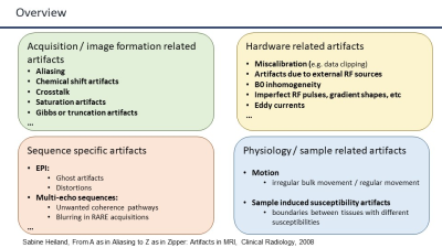
This lecture describes the most common artifacts found on preclinical MRI scanners related to acquisition, image formation, hardware, sequence specific issues and physiological properties. It starts with a brief review of k-space and image formation, then it describes the physical principles of various artifacts, provides examples of preclinical images specifically acquired to show them and discusses different strategies to mitigate artifacts, both at the acquisition and data post-processing level. Some of the artifacts covered in this lecture are aliasing, chemical shift, Gibbs ringing, field inhomogeneities, Eddy currents, ghosts, susceptibility artifacts and motion.
|
|
| Gibbs-Ringing Artifact Removal
Elias Kellner
The gibbs-ringing (also known as "truncation artifact" or "spectral leakage" ) is a result from reconstruction if images from a finite (bounded) k-space. It results in oscillations according to the sinc function in the vicinity of sharp image gradients at tissue boundaries. In this presentation, examples of the artifact are illustrated and simulations are shown to understand it better. Finally, correction strategies are discussed.
|
||
 |
Simulation of Artifacts for Validating Post-Processing Correction Techniques
Gary Zhang
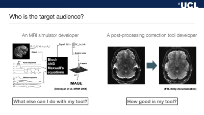
This presentation reviews how simulation of MRI artifacts can help validate post-processing correction techniques. First, typical artifacts that are routinely corrected via post-processing are presented. Second, the strength of validation with MRI simulation over alternative validation approaches is discussed. Finally, a range of practical examples of MRI artifact simulation development and their use for validation are detailed.
|
The International Society for Magnetic Resonance in Medicine is accredited by the Accreditation Council for Continuing Medical Education to provide continuing medical education for physicians.
 Watch the Video