Weekend Course
Imaging Degradation & Inflammation
ISMRM & SMRT Annual Meeting • 15-20 May 2021

Weekend Course
Imaging Degradation & Inflammation
| Concurrent 1 | 15:15 - 16:00 | Moderators: Jorge Davila Acosta & Liliana Ma |
| Inflammatory Demyelinating Disease & Treatment Effects
Matilde Inglese
|
||
 |
Idiopathic Lung Disease & Cystic Fibrosis
Sean Fain
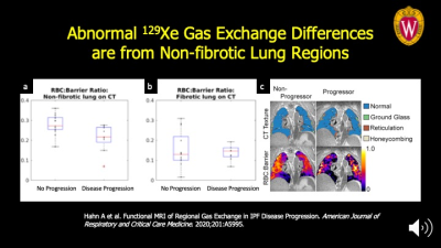
Idiopathic pulmonary fibrosis (IPF) and cystic fibrosis are deadly chronic lung diseases that have recently had breakthroughs in targeted drug therapies. Image-based biomarkers of ventilation and gas exchange using hyperpolarized 129Xe MRI are well suited to sensitively identify patients with progressive disease and monitor response to these new drug therapies. Additionally, ultra-short echo time (UTE) MRI has made significant advances in visualizing clinically relevant disease structures in both IPF and CF and can complement HP 129Xe MRI for longitudinal studies without requiring ionizing radiation. This structure-function imaging capability of MRI makes earlier and more frequent disease assessment feasible.
|
|
 |
Non-Ischemic Cardiomyopathies
Ethan Rowin
Cardiovascular magnetic resonance (CMR) provides comprehensive characterization of the heart in patients with hypertrophic cardiomyopathy (HCM) including precise definition of left ventricular wall thickness and myocardial tissue characterization with late gadolinium enhancement (LGE). CMR contributes to the identification of patients at risk for sudden death including high risk features of LV apical aneurysms and extensive LGE. CMR in conjunction with current management strategies have resulted in a HCM becoming a treatable disease with low mortality rates.
|
|
| Vasculitis
Subha Raman
|
||
| Hepatic Inflammation & Fibrosis-MR Elastography
Frank Miller
Hepatic inflammation and fibrosis are important causes of cirrhosis and hepatocellular carcinoma. The imaging findings of cirrhosis are often difficult to diagnosis on conventional imaging. MR elastography is the best method to assess for increased stiffness associated with fibrosis and inflammation. This talk will discuss the technique, interpretation and performance of MR elastography. Newer techniques including 3D MRE will be discussed. In addition, the patterns of findings on MR elastography will be discussed in relationship to different disease processes. Other causes of elevated stiffness will be demonstrated. Challenges such as iron overload will be discussed.
|
||
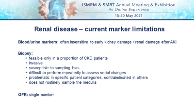 |
Acute & Chronic Kidney Diseases
Anna Caroli
Renal MRI has the potential to overcome the limitations of current renal disease markers and improve kidney disease management in both chronic and acute settings. Renal MRI offers a wide spectrum of techniques that could be combined in a multiparametric approach to gain the best insight into kidney pathophysiology. In this educational presentation, the most widely used renal MRI techniques (DWI, BOLD, ASL, Phase-contrast MRI, T1/T2 mapping) are presented along with their clinical applications and new advances in the field. Current challenges, recent achievements, and a roadmap for clinical translation of renal MRI biomarkers are also discussed.
|
|
 |
Imaging in IBD - a clinical perspective
Tom Watson
MR Enterography forms the mainstay of imaging for inflammatory bowel disease in adults and children. Ultrasound and CT are useful adjuncts in specific circumstances. Assessment of inflammation in IBD requires a multi-sequence approach using fluid-sensitive, diffusion-weighted and contrast enhanced sequences. Currently it is difficult to assess accurately, the relative contributions of inflammation and fibrosis within a given area of disease though this is possible. Newer techniques such as magnetisation transfer factor and quantified dynamic assessment of gut motility offer some promise in these areas.
|
|
| Spondylarthropathy
Chiara Giraudo
This lecture is for radiologists and scientists working in the musculoskeletal field with a special interest in inflammatory rheumatic diseases.The classification of spondyloarthropathies including a brief overview of clinical and laboratory findings will be provided.The optimization of MR protocols according to international guidelines and the main MR signs will be discussed.Moreover, the role of qualitative and quantitative MR imaging including advanced techniques, such as machine learning will be addressed.At the end of this lecture, the audience will have gained new knowledge about the diagnostic process for spondyloarthropathies and the application of basic and advanced MR imaging in this field.
|
The International Society for Magnetic Resonance in Medicine is accredited by the Accreditation Council for Continuing Medical Education to provide continuing medical education for physicians.
 Watch the Video