Digital Poster
Diffusion Modeling, Software, Simulation
ISMRM & ISMRT Annual Meeting & Exhibition • 10-15 May 2025 • Honolulu, Hawai'i

Digital Poster
Diffusion Modeling, Software, Simulation
 |
Computer Number: 65
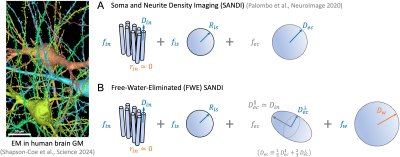
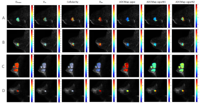
4032. Free-Water-Eliminated
(FWE)-SANDI for improving the accuracy of in vivo apparent soma
and neurite imaging using high-gradient diffusion MRI
H. Lee, K-S Chan, Y. Ma, S. Huang, H-H Lee
Massachusetts General Hospital, Charlestown, United States
Impact: The proposed Free-Water-Eliminated (FWE)-SANDI
model reduces the partial volume effect from free water and
improves the accuracy of non-invasive microstructural
imaging in the brain gray matter, allowing ones to better
study underlying mechanisms of cellular changes in
neurodevelopment and neurodegeneration.
|
|
 |
Computer Number: 66
4033. An
analytical model of restricted diffusion in dendritic spines.
K. Şimşek, M. Jallais, J. Valette, M. Palombo
Cardiff University, Cardiff, United Kingdom
Impact: We developed a biophysical model for
characterizing diffusion MR signal from spiny dendrites,
providing a foundation for designing dMRI/dMRS methods to
non-invasively quantify changes in spine density and
morphology, hallmarks of neuroplasticity, and several
neurodegenerative diseases, and neurological disorders.
|
|
 |
Computer Number: 67
4034. Comparison
of Measures of Axonal Loss in Diffusion Models in Healthy
Controls and Patients with Multiple Sclerosis at 3T
A. Witt, I. Stuart, L. Narisetti, G. Sweeney, K. O'Grady, S.
Smith, S. By, K. Schilling
Vanderbilt University Institute of Imaging Science, Nashville, United States
Impact: Volume fraction measures from novel diffusion
models are sensitive to disease pathology in brain and
spinal cord lesions of patients with multiple sclerosis,
providing evidence of the utility of specific diffusion
measures to capture axonal loss.
|
|
 |
Computer Number: 68
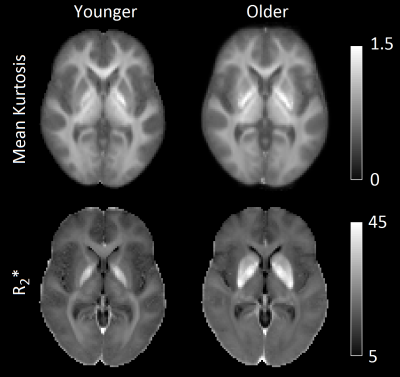
4035. Associations
between iron and mean kurtosis in iron-rich grey matter nuclei
in aging
J. Langley, K. Solis, V. Masjedizadeh, M. Shao, I. Bennett,
X. Hu
University of Calfornia Riverside, Riverside, United States
Impact: Our findings indicate that higher mean kurtosis
in iron-rich grey matter structures may be due to decreases
in signal-to-noise ratios from iron deposition.
|
|
 |
Computer Number: 69
4036. Predicting
Mesoscopic Larmor Frequency Shifts in White Matter with
Diffusion MRI - A Monte-Carlo Study in Axonal Phantoms
A. Sandgaard, S. Jespersen
Aarhus University, Aarhus, Denmark
Impact: µQSM may improve estimation of tissue magnetic
susceptibility and lead to susceptibility imaging with
higher diagnostic value.
|
|
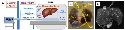 |
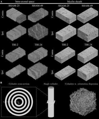
Computer Number: 70
4037. Validation
of Intravoxel Incoherent Motion MRI using Perfused Explanted
Human Livers
G. Simchick, J. Rice, L. Gober, D. Rice, J. Philip, A.
Roldan-Alzate, D. Hernando
University of Wisconsin-Madison, Madison, United States
Impact: Perfused explanted human livers may serve as
biologically accurate systems for validating quantitative
IVIM techniques. Importantly, perfused explanted livers
contain two distinct compartments (i.e., diffusion and
perfusion components of the IVIM signal were independent and
dependent on flow rate, respectively).
|
|
 |
Computer Number: 71
4038. Diagnostic
Value of Using Time-Dependent Diffusion MRI to Identify Benign
and Malignant Breast Tumors
L. Bao, T. Ji, Z. Wang, Y. Sun, Y. Qiu, Z. Shen, X. Zhang
The First Hospital of Jilin University , changchun, China
Impact: The study's findings could improve diagnostic
strategies for clinicians, enhancing early breast cancer
detection and treatment planning. It paves the way for
further research on TD-MRI applications in other cancer
types, ultimately benefiting patient outcomes and overall
care.
|
|
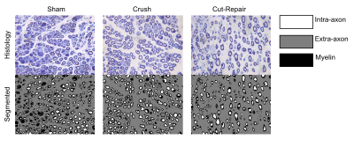 |
Computer Number: 72
4039. Numerical
Validation of Multi-Compartment Diffusion Biomarkers of
Peripheral Nerve Trauma
T. Ketsiri, K. Chen, J. Xu, R. Dortch
Barrow Neurological Institute, Phoenix, United States
Impact: The spherical mean technique (SMT), a
multi-compartmental diffusion MRI model, demonstrated
potential as a biomarker of peripheral nerve regeneration
following injury and surgical repair.
|
|
 |
Computer Number: 73
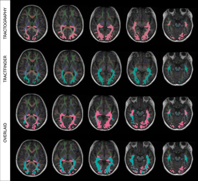
4040. Tractfinder
for paediatric optic radiation segmentation
Y. J. Li, K. Seunarine, J. Clayden, C. Clark
University College London, London, United Kingdom
Impact:
Manual tasks and requirement for user-expertise in tractography pipelines hinder utilisation in clinical settings. We show Tractfinder’s potential for fast, reliable tract segmentation in paediatric patients and control datasets, reducing workload and eliminating the need for specialist tractography training. |
|
 |
Computer Number: 74
4041. Rotation-Free
Estimation of Anisotropic Transverse Relaxation & Larmor
Frequency with Diffusion MRI: A Monte Carlo Study in Axonal
Phantoms
A. Sandgaard, S. Jespersen
Aarhus University, Aarhus, Denmark
Impact: Modeling a multi-spin-echo diffusion MRI signal
with delayed read-outs per echo enables estimation of
orientation-dependent susceptibility parameters without
imaging multiple sample orientations. This approach could
improve sensitivity to tissue characteristics, aiding
studies of myelination and other white matter features.
|
|
 |
Computer Number: 75
4042. Histologically
Informed Periodic Axon Substrate Generator for Time-Dependent
Diffusion MRI
T. Nguyen, M. Does, K. Harkins
Vanderbilt University, Nashville, United States
Impact:
The framework developed can facilitate more accurate Monte Carlo simulations without confounding effects of boundary artifacts for investigating microstructural contributions to diffusion MRI. |
|
 |
Computer Number: 76
4043. MyCaliber:
Axon diameter mapping from myelin water diffusion -- Theory,
Resolution Limit, and Monte Carlo simulations
H. H. Lee, D. Novikov, E. Fieremans, S. Huang
Athinoula A. Martinos Center for Biomedical Imaging, Charlestown, United States
Impact: Measuring restricted diffusion of myelin water
in-between myelin sheaths using diffusion MRI enables to in
vivo measure axon radius. Simulation results demonstrated
its applicability at SNR≥5 on Connectome 2.0 scanner. The
protocol can be adapted for clinically available
high-gradient-performance scanners.
|
|
 |
Computer Number: 77
4044. Validation
of MRI diffusion models using synchrotron radiation imaging in
custom microstructural phantoms
A. Maiuro, M. Fratini, L. Massimi, S. Cipiccia, S. Marathe,
D. Batey, F. Zhou, G. Parker, S. Capuani, M. Palombo
Sapienza University of Rome, Rome, Italy
Impact:
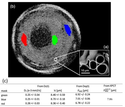
A multi-scale approach based on XPCT has been proposed for validating dMRI models applied on DW data obtained on brain-mimicking phantoms. The agreement between dMRI and XPCTresults pave the way for new possibilities of dMRI validation in ex-vivo brain samples. |
|
 |
Computer Number: 78
4045. Diffusion
MRI-based Explicit Microstructural Imaging Analysis
F. Liu, Z. Wang, L. Chen, W. Zhong, J. Xu, H. Guo, D. Shi
Center for Biomedical Imaging Research, School of Biomedical Engineering, Tsinghua University, Beijing, China
Impact: Clinical applications of cell size imaging are
limited by current implicit model fitting, which is
time-consuming and poorly reproducible. We developed an
explicit imaging method by designing diffusion-weighted
sequences, improving unstable fitting and shortening
computational time.
|
|
 |
Computer Number: 79
4046. Histology
Validation of Generalized Diffusion Basis Spectrum Imaging in
Postmortem Alzheimer Disease Brain
M. Jiang, W. Wu, Y. Nan, A. Patel, E. Franklin, R. Perrin,
T. Benzinger, Y. Wang, Q. Wang
Washington University School of Medicine, Saint Louis, United States
Impact: g-DBSI could enable earlier, non-invasive
monitoring of AD progression, offering a valuable tool for
studying neuroinflammation and cellular changes directly in
gray matter.
|
|
 |
Computer Number: 80
4047. Multi-echo
NODDI with released intrinsic diffusivity in the healthy human
brain tissue
E. Farrher, K-H Cho, R. Buschbeck, C-W Chiang, S-M Huang,
M-J Chen, C-H Choi, L-W Kuo, N. J. Shah
Forschungszentrum Jülich, Jülich, Germany
Impact: Incorporating an l2-norm
regularisation term into the estimation of MTE-NODDI
parameters allows the intrinsic diffusivity to be released
whilst ensuring fitting stability. This opens the
possibility of using MTE-NODDI to investigate a great
variety of tissue pathologies.
|
 Back to Meeting Home
 Back to the Program-at-a-Glance
The International Society for Magnetic Resonance in Medicine is accredited by the Accreditation Council for Continuing Medical Education to provide continuing medical education for physicians.
 View
Presentation Video
View
Presentation Video