Digital Poster
Diffusion Microstructure 2
ISMRM & ISMRT Annual Meeting & Exhibition • 10-15 May 2025 • Honolulu, Hawai'i

 |
Computer Number: 81
3891. Classification
of Benign Ovarian Lesions and Epithelial Ovarian Cancer Subtypes
Using MRI Cytometry-Derived Microstructural Parameters
W. Yue, R. Han, D. Zheng, H. Li, Q. Yang
Beijing Chaoyang Hospital, Capital Medical University, Beijing, China
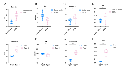
Impact: For the first time, MRI cytometry demonstrated
high feasibility and diagnostic accuracy for non-invasively
distinguishing benign ovarian lesions from epithelial
ovarian cancers (EOCs) and further differentiating between
type I and II EOCs.
|
|
 |
Computer Number: 82
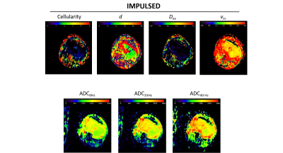
3892. Imaging
Microstructural Parameters of Benign and Malignant Breast Tumor
in Patient Using Temporal Diffusion Spectroscopy
S. Peng, P. Sun, J. Tao, J. Liu, F. Yang
Wuhan Union Hospital, Wuhan, China
Impact: The time-dependent diffusion MRI parameters have
the potential to evaluate the microstructural
characteristics of breast lesions and serves as a
non-invasive tool to probe tumor pathologies in both benign
and malignant breast lesions.
|
|
 |
Computer Number: 83
3893. Utility
of OGSE-Based Microstructural Parameters in Predicting Lymph
Node Metastasis in Breast Cancer
J. Bai, S. Chen, M. Chen, X. Guo, Z. Xiao, F. Thorsten, J.
Shu
The Affiliated Hospital of Southwest Medical University, Luzhou, China
Impact: This study demonstrates that OGSE-based
microstructural MRI parameters provide a reliable,
non-invasive tool for assessing lymph node metastasis in
breast cancer. These findings could enhance diagnostic
accuracy, guide surgical decisions, and improve patient
outcomes.
|
|
 |
Computer Number: 84
3894. Multi-Shell
Diffusion MRI Reveals Effects of Extracranial Carotid Artery
Disease on Brain Microstructure
H. Wiskoski, J. Arias, L. Do, A. Pugazhendhi, R. Mushtaq, K.
Johnson, M. Altbach, T. Trouard, C. Weinkauf
The University of Arizona, Tucson, United States
Impact: This study's findings provide a foundation for
using diffusion MRI to identify early brain changes in
patients with carotid artery disease, potentially enabling
proactive interventions. Future research may explore
targeted therapies to mitigate neurodegeneration linked to
vascular disease.
|
|
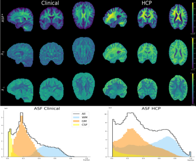 |
Computer Number: 85
3895. Clinically
feasible axonal fraction imaging
T. Thøgersen, T. Dyrby, M. Pizzolato
Technical University of Denmark, Kgs. Lyngby, Denmark
Impact: The ASF is related to the volume occupancy of
axons and could be used to characterize pathology. We enable
its estimation using conventional diffusion MRI data from a
clinical scanner, while at the same time minimizing model
assumptions and degeneracy.
|
|
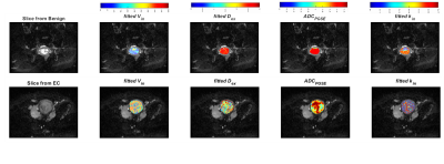 |
Computer Number: 86
3896. TIME-DEPENDANT
DIFFUSION-WEIGHTED MRI IN MAPPING TUMOUR MICROENVIRONMENT IN
ENDOMETRIAL CARCINOMA.
E. Lee, D. Shi, R. Singh, J. Zhang, A. Hwang, F. Liu, Z.
Wang, T. Feiweier, J. Wei, H. Guo
University of Hong Kong, Hong Kong, China
Impact: Time-dependent diffusion-weighted MRI allows
non-invasive characterisation of the tumour microenvironment
of endometrial carcinoma, deepening our understanding of the
complex interplay between tumour and its microenvironment,
providing opportunities for identification of drug targets
and monitoring disease evolution throughout treatment.
|
|
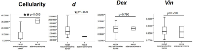 |
Computer Number: 87
3897. Microstructural
mapping based on time-dependent diffusion MRI for
differentiating rectal neuroendocrine tumor and rectal
adenocarcinoma
Y. Li, X. Chen, X. Dong, S. Yi, M. Chen, P. Zhou
Sichuan Clinical Research Center for Cancer, Sichuan Cancer Hospital & Institute, Sichuan Cancer Center, Affiliated Cancer Hospital of University of Electronic Science and Technology of China, Chengdu, China
Impact: Microstructural parameters derived from td-dMRI
have the potential to serve as imaging biomarkers for the
non-invasive differentiation of rectal neuroendocrine
tumors, thus aiding in the prediction of histopathological
types of rectal cancer.
|
|
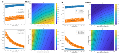 |
Computer Number: 88
3898. Detecting
dendritic spine density with double diffusion encoding magnetic
resonance spectroscopy
M. Jallais, S. Malaquin, K. Simsek, J. Valette, M. Palombo
Cardiff University, Cardiff, United Kingdom
Impact: Using numerical simulations and in-vivo mouse
experiments we showed that DDE-MRS may be a promising
non-invasive method for estimating dendritic spine density
in-vivo, providing a new avenue for in-vivo studies of
healthy and pathological brain gray matter microstructure.
|
|
 |
Computer Number: 89
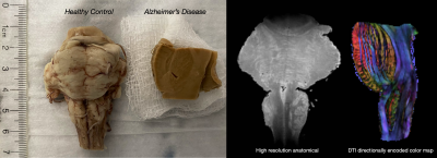
3899. Double
diffusion encoded MRI to identify Alzheimer’s disease pathology
in postmortem brainstem by Diffusion Tensor Subspace Imaging
(DiTSI)
C. Comrie, V. Sandrin, L. Dieckhaus, G. Serrano, T. Beach,
M. Bondi, S. Solders, V. Galinsky, L. Frank, E. Hutchinson
University of Arizona, Tucson, United States
Impact: If double diffusion encoding MRI can be
optimized for the detection of specific pathology in
Alzheimer's disease and other brain disorders, a new class
of improved imaging markers may be possible.
|
|
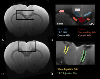 |
Computer Number: 90
3900. Are
the Effects Resulting from an LPC-Induced Focal Lesion in the
White Matter Truly Localised?
E. Thomson, M. Corral-Bolaños, T. Dyrby
Danish Research Centre for Magnetic Resonance, Hvidovre, Denmark
Impact: This study suggests that no significant response
is detected surrounding the LPC injection site nor the
surrounding cortex, indicating that this model can be
trusted for electrophysiology and expected conduction
velocity changes are related only to the LPC-induced lesion
area.
|
|
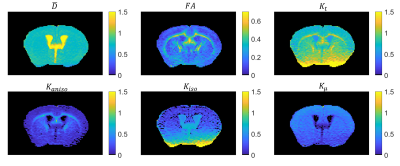 |
Computer Number: 91
3901. Correlation
Tensor MRI of the Mouse Brain at 3 Tesla
R. Henriques, A. Ianus, N. Shemesh, R. Simões
Champalimaud Foundation, Lisbon, Portugal
Impact: This study reports the first implementation of
Correlation Tensor MRI (CTI) on a 3T preclinical MRI
scanner. This could facilitate the translation of CTI’s
microstructural insights in animal models of human disease
to clinical applications in patients.
|
|
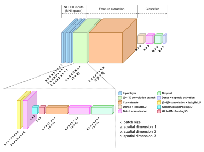 |
Computer Number: 92
3902. Prediction
of amyloid positivity based on white matter structural integrity
in non-demented individuals using 3D CNN
P. Pattiam Giriprakash, Z. Yang, D. Cordes, A. Bender
Cleveland Clinic, Las Vegas, United States
Impact:
Early detection of elevated amyloid burden in non-demented patients using diffusion MRI can stratify patients with a high risk of developing Alzheimer’s disease. It could potentially serve a prognostic biomarker for recommending early clinical intervention. |
|
 |
Computer Number: 93
3903. Application
Research of Cellular Microstructural Parameters Based on MRI in
Predicting Lymph Node Metastasis and Tumor Deposit in Rectal
Cancer
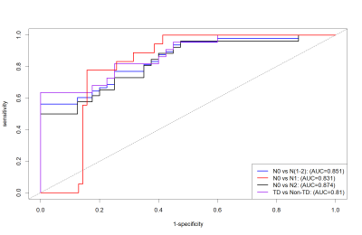
S. Yi, X. Dong, Y. Li, M. Chen, X. Chen
Sichuan Clinical Research Center for Cancer, Sichuan Cancer Hospital & Institute, Sichuan Cancer Center, Affiliated Cancer Hospital of University of Electronic Science and Technology of China, Chengdu, China
Impact: The parameter diameter derived from td-dMRI has
the potential to serve as an imaging biomarker for the
preoperative prediction of LNM and TD.
|
|
 |
Computer Number: 94
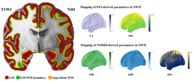
3904. Superficial
white matter microstructural impairments correlate with
functional alterations and disease severity in early-stage ALS
S. Zhuang, H. Chen
Fujian Medical University Union Hospital, Fuzhou , China
Impact: Focusing on SWM evaluation offers a new
perspective on the interaction between structural and
functional disruptions in ALS, highlighting a crucial
neurobiological substrate related to disease progression.
|
|
 |
Computer Number: 95
3905. Prediction
of Ki-67 Expression in Soft Tissue Tumors Using Time-Dependent
Diffusion MRI
Z. Yan, J. Zhao, X. Feng, X. Zhang, L. Duan, M. Wang, H. Yu
The Third Hospital of Hebei Medical University, Shijiazhuang, China
Impact: Time-dependent
diffusion MRI provides a noninvasive method for preoperative
evaluation of malignant proliferation in soft tissue tumors,
offering a promising alternative to traditional diagnostic
techniques.
|
|
 |
Computer Number: 96
3906. Time-dependent
diffusivity is sensitive to pathology in an Alzheimer’s disease
mouse model: a multidimensional MRI pilot study
P. S. Or, M. Yon, O. Narvaez, A. Sierra, D. Topgaard, D.
Benjamini
National Institute on Aging, NIH, Baltimore, United States
Impact: Our findings establish the reliability of
diffusion-time dependent metrics and their sensitivity to AD
pathology, supporting further large-scale studies to explore
the potential of md-MRI for earlier AD diagnosis and
improved prognostic outcomes.
|
 Back to Meeting Home
 Back to the Program-at-a-Glance
The International Society for Magnetic Resonance in Medicine is accredited by the Accreditation Council for Continuing Medical Education to provide continuing medical education for physicians.
 View
Presentation Video
View
Presentation Video