Digital Poster
Bone
ISMRM & ISMRT Annual Meeting & Exhibition • 10-15 May 2025 • Honolulu, Hawai'i

 |
Computer Number: 81
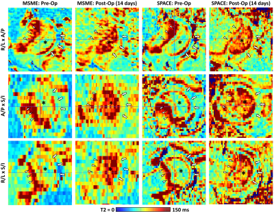
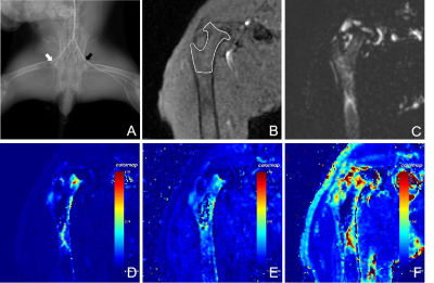
3104. Evaluation
of a 5-minute, clinically feasible T2 mapping sequence to detect
ischemic injury to the developing femoral head: a piglet model
study
C. Johnson, S. Parvaze, N. Nelson, S. Novom, A. Amann, E.
Buko, S. Moeller, A. Armstrong, F. Toth
University of Minnesota, Saint Paul, United States
Impact: Ischemic injury to the femoral head can be
detected using a 5-minute, clinically available T2 mapping
sequence that is suitable for imaging young children. This
technique may facilitate clinical translation of T2 mapping
for detection and monitoring of Legg-Calvé-Perthes disease.
|
|
 |
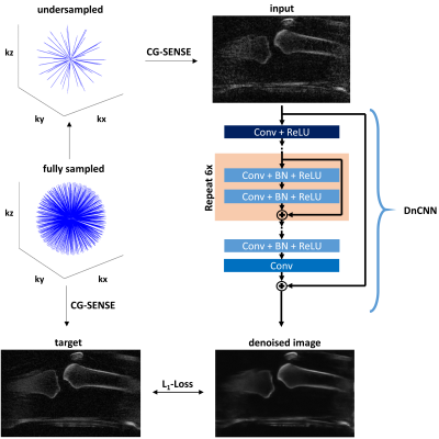
Computer Number: 82
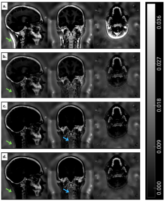
3105. Towards
High Resolution Large Field-of-View Bone MRI
T. Catargiu, T. Wood
King's College London, London, United Kingdom
Impact: Using MRI for bone imaging would remove the
associated risks from the ionizing radiation required for
CT. The developments shown here are a step towards a
robust, high resolution MRI bone scan over a large FOV.
|
|
 |
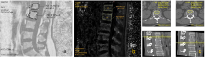
Computer Number: 83
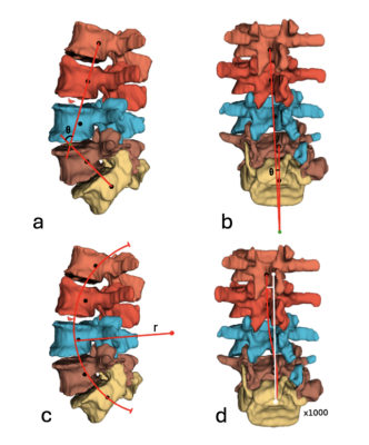
3106. Magnetic
resonance imaging provides comparable spinal curvature
measurements to computerized tomography
J. Miller, K. Marusich, C. Beaulieu, A. Chaudhari, G. Gold
Occidental College, Los Angeles, United States
Impact: This study impacts research done between CT
scans and MR images, supporting intra-modality spinal
curvature calculations from vertebral centroids calculated
semi-automatically. This study also supports the equivalency
of spinal curvature calculations from CT scans, T1-weighted
and T2-weighted MR images.
|
|
 |
Computer Number: 84
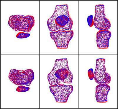
3107. A
Comparison of Two Combined Shape Modeling Approaches of Knee
Bones for Patellofemoral Instability
J. Peters, M. Yang, R. Lartey, C. Winalski, L. Farrow, X. Li
The Cleveland Clinic Foundation, Cleveland, United States
Impact: Better characterization of the shapes and
orientations of skeletal morphologies and their correlations
could lead to improved diagnosis and more targeted
treatments for musculoskeletal pathologies such as PFI.
|
|
 |
Computer Number: 85
3108. Multi-parameter
quantitative MRI study on the intervention of exogenous hydrogen
sulfide in bone micro-environment of diabetic rabbits with CLI
Y. Gao, Y. Zha, W. Liu
Renmin hospital of Wuhan university, Wuhan, China
Impact: This study provides quantitative imaging
evidence for the protective mechanism of H₂S in diabetic
critical limb ischemia complications.
|
|
 |
Computer Number: 86
3109. A
data-driven approach to accelerate IR-prepared UTE Bone Imaging
of the Knee
P. Nunn, O. Schad, H. Huflage, J-P Grunz, T. Bley, J.
Tran-Gia, T. Wech
University Hospital Würzburg, Würzburg, Germany
Impact: The proposed reconstruction technique
demonstrates potential for MR-based assessment of osseous
tissue within clinically appealing scan time.
|
|
 |
Computer Number: 87
3110. Evaluation
of 3.0T MR IDEAL-IQ for fat fraction and T2 * value to diagnose
osteoporosis
Y. Zhang, Z. Li, X. Xiang, Y. Liu, J. Chen
Chengdu Sports University, Chengdu Sichuan, China
Impact: MRI-based FF and T2* value can be used as a
supplement to BMD for early assessment of fracture risk to
prevent fragility fractures.
|
|
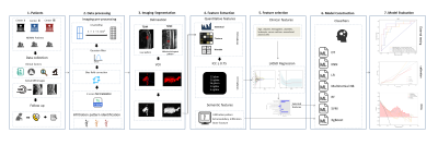 |
Computer Number: 88
3111. A
semantic-enhanced integrated radiomics model based on spinal MRI
for predicting early relapse in multiple myeloma: A multi-center
study
C. Liu, C. Ling, Q. Yang, W. Yang, Y. Zhao
The Third Affiliated Hospital of Southern Medical University (Academy of Orthopedics, Guangdong Province), GuangZhou, China
Impact: Integrating imaging and semantic features
improved model accuracy and interpretability, enabling early
risk identification and supporting personalized MM
treatment.
|
|
 |
Computer Number: 89
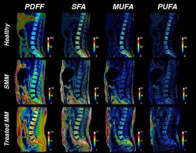
3112. Characterizing
Bone Marrow Adipose Tissue Composition in Multiple Myeloma Using
Chemical Shift Encoded MRI
D. Martel, M. Bruno, M. Mayerhoefer
NYU Langone, New York, United States
Impact: These findings highlight the clinical potential
of CSE-MRI in assessing metabolic biomarkers for MM staging
and treatment monitoring. This study lays the groundwork for
a more precise, non-invasive approach to MM assessment,
warranting further investigation in larger cohorts.
|
|
 |
Computer Number: 90
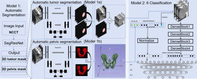
3113. Automated
Sacral Tumor Localization and Multi-Class Identification of Six
Tumor Types in NCCT with an innovative CL-MedImageNet Fusion
Model
F. Zheng, P. Yin, W. Zhang, N. Hong
Peking University people’ hospital, Beijing, China
Impact: Our model reliably predicts sacral tumor types
using a fully automated NCCT process, improving
individualized treatment planning with its high
reproducibility and generalizability.
|
|
 |
Computer Number: 91
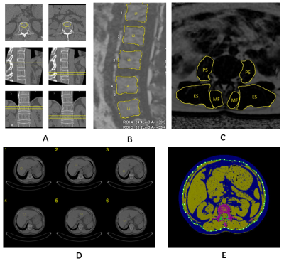
3114. Visceral,
bone marrow, and muscle adiposity have effects on volumetric
bone mineral density in women: a study based on MRI and QCT
Z. Li, X. Xiang, Y. Zhang, H. Liang, J. Chen
Chengdu Sport University, Chengdu, China
Impact: Our study showed that VAT, BM PDFF and PSM PDFF
were negatively correlated with vBMD. The findings elucidate
the role of adipose tissue distribution in osteoporosis and
provide new ideas for clinical diagnosis and treatment of
osteoporosis.
|
|
 |
Computer Number: 92
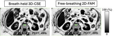
3115. Bone
Marrow Proton Density Fat Fraction Measurement Using Flip-Angle
Modulated Chemical Shift-Encoded MRI
R. do Vale Souza, A. Anagnostopoulos, J. Tang, D. Tamada, J.
Frederik Heidenreich, S. Reeder, D. Hernando, A. Pirasteh
University of Wisconsin-Madison, Madison, United States
Impact: FAM-based CSE MRI is a promising tool for
motion-robust measurement of bone marrow PDFF, which may
help characterization of many hematologic disease processes.
However, it requires further validation and addressing
potential sources of bias.
|
|
 |
Computer Number: 93
3116. Differentiating
multiple myeloma and bone metastasis on spine MRI using
radiomics-based machine learning and deep learning models
L-N Do, S. W. Kang, D. Yun, I. Park
Chonnam National Univeristy, Gwangju, Korea, Republic of
Impact: The results from this study suggest that the
proposed approach may provide a valuable assisting tool in
distinguishing two challenging conditions, MM and metastasis
on spine MRI.
|
|
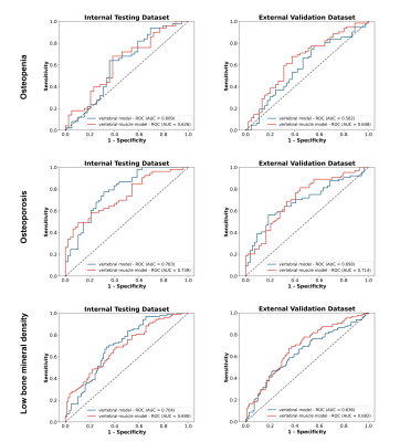 |
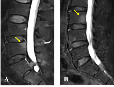
Computer Number: 94
3117. MRI-Based
Deep Learning for Predicting Vertebral Fractures Risk in
Patients with Low Bone Mass: a Multicenter Validation Study
(n=1182)
Y. Yang, T. Zhao, Q. Qiu, Q. Xie, X. Zhang, J. Luo, Z.
Zhang, Y. Wang, J. Wu, C. Huang, X. Zhang
The Third Affiliated Hospital, Southern Medical University, Guangzhou, China
Impact: The paraspinal muscles, as one of the key
structures in maintaining spinal stability, work
synergistically with the vertebrae in predicting vertebral
fracture risk, especially in osteopenia patients.
|
|
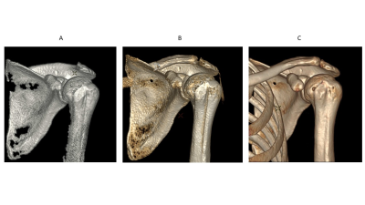 |
Computer Number: 95
3118. 1-Click
Bone Volume Rendering with ZTE MRI
M. Carl, L. Carretero-Gomez, S. Mandava, M. Fung
GE Healthcare, Fallbrook, United States
Impact: Our work has the potential to become a
one-stop-shop imaging approach with the ability to get
co-registered bone and soft-tissue images in a single,
radiation-free MR exam.
|
|
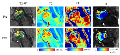 |
Computer Number: 96
3119. Quantitative
MRI Techniques to Monitor Changes in Osteonecrosis of the
Femoral Head Following Core Decompression: A Preliminary Study
E. Buko, E. Cheng, J. Ellermann, C. Johnson
University of Minnesota, Saint Paul, United States
Impact: Quantitative MRI techniques are sensitive in
detecting compositional changes in ONFH lesions following
core decompression treatment. These methods may help
identify patients who can benefit from a second intervention
and help improve patient management and optimize therapeutic
approaches for ONFH.
|
 Back to Meeting Home
 Back to the Program-at-a-Glance
The International Society for Magnetic Resonance in Medicine is accredited by the Accreditation Council for Continuing Medical Education to provide continuing medical education for physicians.
 View
Presentation Video
View
Presentation Video