Digital Poster
Pulse Sequences for Body & Cardiovascular Imaging
ISMRM & ISMRT Annual Meeting & Exhibition • 10-15 May 2025 • Honolulu, Hawai'i

Digital Poster
Pulse Sequences for Body & Cardiovascular Imaging
 |
Computer Number: 33
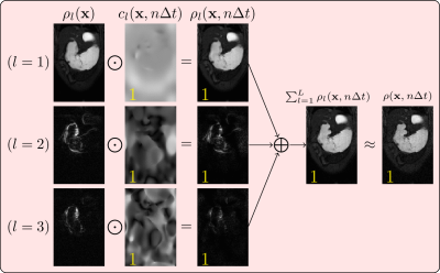
2610. Spatiotemporal
maps for dynamic MRI reconstruction: a proof-of-principle
demonstration on single-coil animal gastrointestinal data

R. Lobos, X. Wang, Z. Liu, J. Fessler, D. Noll
University of Michigan, Ann Arbor, United States
Impact: Spatiotemporal maps provide a parsimonious
voxel-dependent expansion for the dynamic MRI signal, even
when voxels present various temporal/spectral
characteristics. They can be efficiently calculated from
autocalibration data, and can be synergistically combined
with modern regularizers to reconstruct highly accelerated
data.
|
|
 |
Computer Number: 34
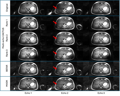
2611. Automated
streaking artifact suppression for multi-echo radial abdominal
data compatible with advanced reconstruction techniques
B. Toner, S-F Shih, F. Han, E. Ahanonu, U. Goerke, K.
Johnson, H. Wu, M. Altbach, A. Bilgin
The University of Arizona, Tucson, United States
Impact:
The proposed destreaking method was demonstrated to be equivalent when applied to multi-coil images, multi-coil k-space, or coil sensitivity maps, providing flexibility when being incorporated into existing reconstruction algorithms. It out-performed existing techniques and improved the reconstruction of multi-echo data. |
|
 |
Computer Number: 35
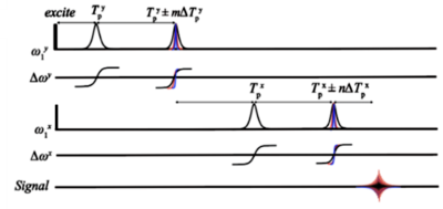
2612. Achieving
2D sub-millimeter images using Phase-Encoded Frequency-modulated
Rabi Encoded Echoes in a B0 gradientless inhomogeneous 0.5T
magnet
D. Pizetta, M. Mullen, P. Jenkins, E. Vidoto, M. Martins, A.
Tannús, M. Garwood, E. Torres
Centro de Imagens e Espectroscopia por Ressonância Magnética - CIERMag - São Carlos Physics Institute, University of São Paulo – IFSC-USP, São Carlos, Brazil
Impact: 2D FREE’s results highlight the potential to
enable high-resolution imaging despite large magnetic field
inhomogeneities and no B0-gradient
hardware. These advancements could enable new low-cost
architectures that are effective and affordable, making MRI
more accessible to the world.
|
|
 |
Computer Number: 36
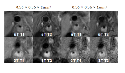
2613. Initial
Experience of Carotid Artery MR Vessel Wall imaging at 5.0T
Q. Wang, X. Zhao, H. Qiao, N. Xu, D. Meng
Center for Biomedical Imaging Research, School of Biomedical Engineering, Tsinghua University, Beijing, China, Beijing, China
Impact: This study demonstrates that 5.0T MR imaging
significantly enhances carotid plaque visualization,
potentially aiding clinicians in early stroke risk
assessment. The findings facilitate future studies on
ultra-high field MRI’s role in diagnosing vulnerable plaque,
advancing preventative stroke care.
|
|
 |
Computer Number: 37
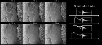
2614. Definition
of the echo time for UTE imaging and short rectangular rf-pulses
M. Krämer, K-H Herrmann, J. Reichenbach
Jena University Hospital - Friedrich Schiller University Jena, Jena, Germany
Impact: Results indicate that defining TE from the
center of short rectangular RF pulses is more accurate,
suggesting the community should adopt this TE definition for
future UTE applications.
|
|
 |
Computer Number: 38
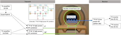
2615. Interleaved
2H and 31P MRSI acquisitions of the liver at 7T employing a
double tuned transmit bore coil and receive body array
L. Stam, M. Konig, M. Gosselink, H. Hoogduin, C. Alborahal,
D. Welting, J. Wijnen, D. Klomp
University Medical Center Utrecht, Utrecht, Netherlands
Impact:
Lossless RF filters enable interleaved 31P and 2H MRSI acquisitions employing a double tuned bore coil and body receive array. This results in substantially improved SNR of metabolite signals from both nuclei, obtained in less examination time. |
|
 |
Computer Number: 39
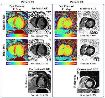
2616. High
resolution synthetic late gadolinium enhancement based on
accelerated post-contrast T1 mapping
J. Gao, Y. Gong, Z. Chen, H. Chen, Y. Emu, X-Y Zhang, Z.
Zhou, W. Jin, S. Hua, C. Hu
Shanghai Jiao Tong University, Shanghai, China
Impact: The proposed method can achieve a similar
accuracy in assessing myocardial scar relative to
conventional LGE, while offering superior image quality,
improved myocardial nulling, and an ability to
simultaneously detect focal and diffuse fibrosis.
|
|
 |
Computer Number: 40
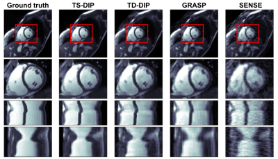
2617. Multi-Slice
Single-Breath-Hold Cardiac CINE with Slice and Time-Dependent
Deep Image Prior at 1.5T and 0.55T
R. De la Sotta, T. Catalán, F. Sahli-Costabal, R. Botnar, C.
Prieto
Millennium Institute for Intelligent Healthcare Engineering, Santiago, Chile
Impact: The proposed approach enables the acquisition of
multiple cardiac CINE slices in just one breath hold, both
at 1.5T and 0.55T. This reduces acquisition time and thus
minimizes slice misalignment for cardiac CINE exams.
|
|
 |
Computer Number: 41
2618. Breathing-speed
quantitative motion imaging using radial acquisition and
motion-subspace reconstruction
V. Murray, R. Otazo
Memorial Sloan Kettering Cancer Center, New York City, United States
Impact: Direct measurement of motion at the speed of
breathing can enable the extraction of functional
information and improve motion characterization for
radiotherapy planning.
|
|
 |
Computer Number: 42
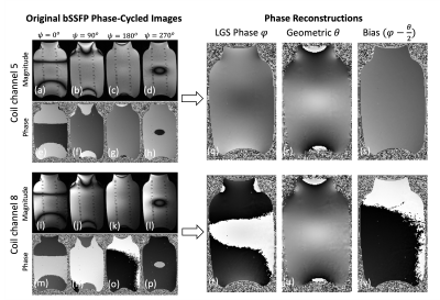
2619. Unraveling
bSSFP Phase: High Quality Field and Bias Mapping
Y. Dong, Q-S Xiang, M. Hoff
University of Washington, Seattle, United States
Impact: The proposed method leverages bSSFP's SNR
efficiency to generate B0 and RF receive-channel bias maps
with high-SNR, smooth variation, and full FOV coverage. This
enables advanced phase-sensitive reconstructions and
correction of phase-related system errors without requiring
additional scans.
|
|
 |
Computer Number: 43
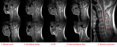
2620. Fast
3D bSSFP Spine Imaging at 0.05 T
J. Hu, Y. Ding, S. Su, V. Lau, J. Zhang, C. Man, A. Leong,
Y. Zhao, E. Wu
The University of Hong Kong, Hong Kong, China
Impact: An optimized bSSFP protocol at 0.05 T was
successfully implemented and achieved fast C- and L-spine
imaging within 4 min. This highlighted the potential of
applying high-quality and fast ULF imaging in clinics.
|
|
 |
Computer Number: 44
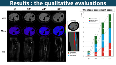
2621. Active
Utilization of the Magic Angle Effect to Enhance Visualization
of Upper Limb Peripheral Nerves
R. Kurosawa, H. Yokota, T. Sada, K. Nitta, I. Nakanishi, H.
Sato, K. Matsumoto, N. Takashi, Y. Masami, T. Iimori, T. Uno
Department of Radiology, Chiba University Hospital, Chiba , Japan
Impact: The magic angle effect enhanced neurography in
qDESS improved the visualization of nerves, which has the
potential to aid in diagnosing and monitoring neuropathy.
|
|
 |
Computer Number: 45
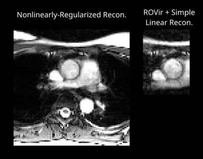
2622. ROVir
Enables Substantially Easier Real-Time Imaging of Small
Regions-of-Interest
C-C Chan, D. Kara, D. Kwon, E. Roselli, C. Nguyen, J. Haldar
University of Southern California, Los Angeles, United States
Impact: High-resolution real-time MRI over a large FOV
is usually highly-undersampled and requires
advanced/time-consuming reconstruction methods. We
demonstrate that ROVir can be used to shrink the size of the
FOV to enable a much simpler/easier reconstruction problem.
|
|
 |
Computer Number: 46
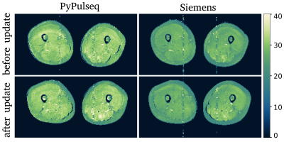
2623. A
standardized open-source MESE sequence implemented in PyPulseq
for reproducible water T2 quantification in skeletal muscle
J. Schäper, F. Santini, C. Weidensteiner
University of Basel, Basel, Switzerland
Impact: The developed vendor-independent MESE sequence
offers the advantage of better transferability between
different scanners. This is an important step towards better
reproducibility of muscle wT2 values in patients with muscle
dystrophies.
|
|
 |
Computer Number: 47
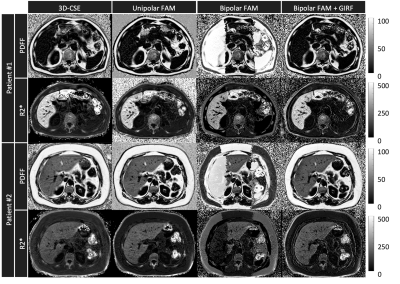
2624. Bipolar
Flip Angle Modulated Chemical Shift Encoded Imaging Enabled with
Gradient Impulse Response Function
J. Wang, J. Tang, D. Tamada, T. Cashen, A. Pirasteh, D.
Hernando, A. McMillan
University of Wisconsin-Madison, Madison, United States
Impact: This study explores the application of GIRF to
enable bipolar 2D-FAM for PDFF quantification. Results are
comparable to conventional 3D-CSE and unipolar 2D-FAM, but
yield scan time reduction, increase in R2* dynamic range,
and shorter ∆TE.
|
|
 |
Computer Number: 48
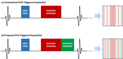
2625. Rethinking
Outer Volume Removal for Fast ECG-Triggered Anatomical Cardiac
MRI
M. Gulle, S. Weingartner, C. Shenoy, M. Akcakaya
University of Minnesota, Minneapolis, United States
Impact: This work proposes a novel pulse sequence for
ECG-triggered CMR sequences, along with a complementary
outer volume removal approach. This simplifies the
reconstruction process, facilitating higher acceleration
rates high-resolution anatomical CMR with reduced artifacts
and improved image clarity.
|
 Back to Meeting Home
 Back to the Program-at-a-Glance
The International Society for Magnetic Resonance in Medicine is accredited by the Accreditation Council for Continuing Medical Education to provide continuing medical education for physicians.
 View
Presentation Video
View
Presentation Video