Digital Poster
Brain Tumors: Metabolism, Spectroscopy & CEST
ISMRM & ISMRT Annual Meeting & Exhibition • 10-15 May 2025 • Honolulu, Hawai'i

Digital Poster
Brain Tumors: Metabolism, Spectroscopy & CEST
 |
Computer Number: 129
2389. Quantitative
Analysis of Dynamic CEST Images to Evaluate Pseudoresponse of
Bevacizumab Treatment
K. Chai, P. Wang, J. Wu, Z. Zhang, K. Schreck, M. Holdhoff,
D. Kamson, L. Blair, J. Laterra, J. Zhou, S. Jiang
Johns Hopkins University, Baltimore, United States
Impact: This study provides a practical approach for
robust and accurate evaluation of Bevacizumab treatment,
demonstrating the superiority of CEST imaging in identifying
pseudoresponse. It shows potential to complement existing
clinical protocols, enabling more precise diagnoses for
glioma patients.
|
|
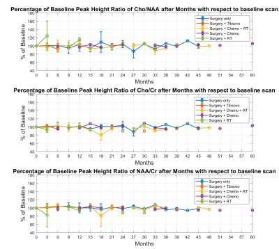 |
Computer Number: 130
2390. Longitudinal
assessment of changes in brain metabolism in the
normal-appearing white matter of patients with lower-grade
glioma.
Y. Liu, T. Luks, A. Autry, S. Vaziri, L. Cheung, J. Lupo, N.
A. Oberheim Bush, S. Chang, J. Villanueva-Meyer, Y. Li
UCSF, San Francisco, United States
Impact: Results of this study can provide insight into
if different treatments can impact a patient’s
neurocognitive function that is tied with metabolite
profile. The finding can also provide suggestions on when
re-treatment is recommended for patients with LrGG.
|
|
 |
Computer Number: 131
2391. A
novel approach to the metabolic characterisation of brain
tumours: edema-corrected OEF. A hybrid MR-PET study.
A-M Oros-Peusquens*, J. Cho, F. Boers, I. Fazli Jaliseh, K-J
Langen, N. J. Shah
Forschungszentrum Jülich, Jülich, Germany
Impact: A simple and fast acquisition method enables
quantitative and metabolic characterisation of brain tumours
and can be implemented in large and multicentre patient
studies. Applying edema correction makes the properties of
underlying tissue accessible, potentially enabling more
precise tumour characterisation.
|
|
 |
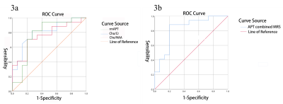
Computer Number: 132
2392. Quantitative
metrics derived from amide proton transfer imaging have improved
performance in evaluating glioma subtypes
H. Zhu, X. Zhang, N. Zheng, W. Zhu
Tongji Hospital, Tongji Medical College, Huazhong University of Science and Technology, Wuhan, China
Impact: The study demonstrates that advanced
quantitative metrics derived from APTw imaging have improved
ability to diagnose glioma, which may serve as useful
imaging biomarkers for glioma evaluation, and facilitate the
clinical management and prognosis of glioma patients.
|
|
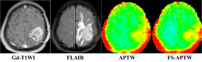 |
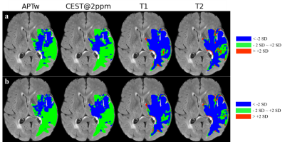
Computer Number: 133
2393. Fluid
suppressed amide proton transfer weighted imaging for
distinguishing true progression from treatment response in
post-treatment gliomas
C-Q Su, J-W Hou, X-C Zhao, S-S Lu
The First Affiliated Hospital of Nanjing Medical University, Nanjing, China
Impact:
FS-APTW imaging offers a more specific characterization of true progression versus treatment response in post-treatment gliomas than APTW imaging. |
|
 |
Computer Number: 134
2394. Initial
experience of characterizing recurrent IDH-mutant astrocytoma
using multi-parametric 1H/HP-13C MRI and histopathological
analysis
X. A. Xing, A. Autry, T. Luks, M. LaFontaine, D. Nair, J.
Lupo, J. Phillips, J. Villanueva-Meyer, H-J Shawn, S. Chang,
Y. Li
UCSF, San Francisco, United States
Impact: This study advances the understanding of glioma
metabolism through the integration of novel
multi-parametric 1H/HP-13C
MRI and tumor histopathological analysis. The insights can
enhance precision in characterizing glioma, potentially
guiding tailored treatment strategies.
|
|
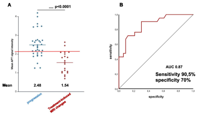 |
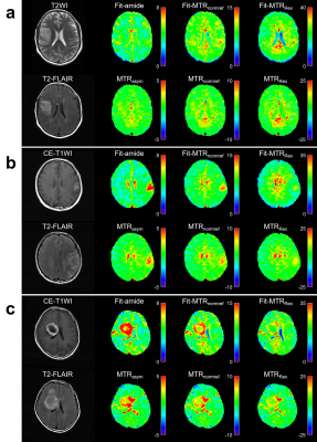
Computer Number: 135
2395. Amide
proton transfer weighted (APTw) imaging for identifying
treatment-induced MRI changes in IDH-wildtype glioblastoma at 3
Tesla
I. Krause, T. Zeyen, A. Decker, F. Kroh, S. Regnery, N.
Schaefer, J. Weller, J. Keupp, C. Katemann, A. Radbruch, U.
Herrlinger, D. Paech
University Hospital Bonn, Bonn, Germany
Impact: APTw imaging shows promise for clinical use in
glioblastoma assessment, particularly for identifying
treatment-induced MRI changes. Future prospective studies
should evaluate the combination of APTw imaging with other
techniques (e.g., DWI and/or perfusion MRI) to further
enhance diagnostic accuracy.
|
|
 |
Computer Number: 136
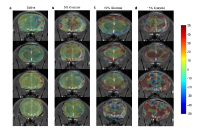
2396. Exploration
of the Boundaries of Glioma through T1ρ-weighted Dynamic
Glucose-enhanced MR Imaging
B. Yin, Y. Zhu, X. Gu, D. Xia, X. Li, Y. Zhao, N. Mei, X.
Liu, P-Y Wu, Y. Lu
Huashan Hospital, Fudan University, Shanghai, China
Impact: T1ρ-DGE MRI acted as a non-invasive visualized
imaging tool for the identification of glioma boundaries.
T1ρ-DGE MRI holds considerable potential for tumor margin,
infiltration, and metabolism of glioma, as well as
therapeutic strategies.
|
|
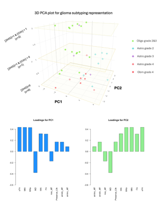 |
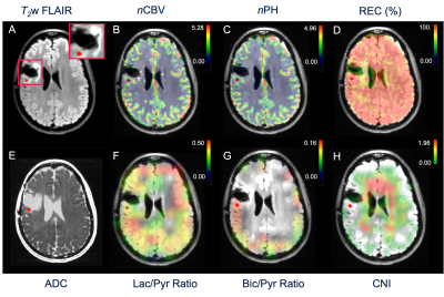
Computer Number: 137
2397. Noninvasive
glioma stratification by multiparametric metabolic and
microstructural MRI at 3T
C. CADIN, M. DIDIER, L. NICHELLI, B. MATHON, F.
SZCZEPANKIEWICZ, M. NILSSON, S. CASAGRANDA, M. ZAISS, F-X
LEJEUNE, S. LEHERICY, M. SANSON, F. BIELLE, M. MARJANSKA, F.
BRANZOLI
Paris Brain Institute - ICM, Inserm U1127, CNRS UMR 7225, Sorbonne Université, UMR S 1127, Equipe labellisée par la Ligue Nationale contre le Cancer, Paris , France
Impact: Characterization of novel biomarkers of glioma
metabolism and microstructure could enhance diagnostics,
deepen understanding of glioma biology, and support an
improved glioma stratification. Our PCA model demonstrated
high accuracy in distinguishing glioma subtypes defined by
WHO 2021 histomolecular classification.
|
|
 |
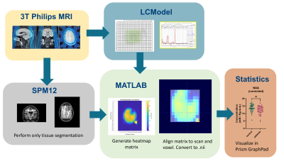
Computer Number: 138
2398. Improved
access and harmonized inline process for 3D spectroscopic MRI to
facilitate clinical integration
V. Khalilzad Sharghi, S. Ahn, A. Trivedi, S. Sheriff, R.
Guo, K. Chow, A. Maudsley, J. Alger, H. Shim, B. Soher
Siemens-Healthineers, Atlanta, United States
Impact: Our work brings high-resolution, 3D metabolic
imaging into clinical practice for glioblastoma, providing
better tools for treatment planning. This advancement could
lead to more effective therapies and new research
opportunities in brain tumor management, benefiting patients
with improved care options.
|
|
 |
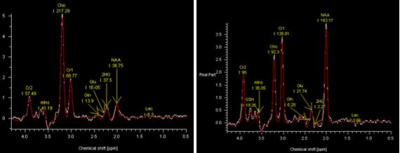
Computer Number: 139
2399. Application
of APT and MRS in the Grading of Brain Gliomas
L. Liu, S. Wan, K. Ai, Y. Zhu, X. Liao
Affiliated Hospital of GuiZhou Medical University, Guiyang, China
Impact: APT and MRS can predict glioma grade
preoperatively. This study suggests that APT and MRS can be
used to predict glioma grade preoperatively, providing
clinicians with important information for planning treatment
strategies and assessing prognosis.
|
|
 |
Computer Number: 140
2400. Towards
fully in-line single-voxel MRI spectroscopy for 2HG
quantification as an IDH biomarker – can it eventually reach
clinical use?
A. Walls, B. Crouch, S. Withey, M-S To, C. Raven, S.
Poonnoose, M. Agzarian, A. Dwyer
South Australian Health and Medical Institute, Adelaide, Australia
Impact: 2HG-MRS achieved with routinely available
acquisition in clinical workflow and translated to inline
analysis software could promote 2HG as a usable biomarker
for IDH status in practice.
|
|
 |
Computer Number: 141
2401. 7T
6,6’-[2H2]-glucose DMI to profile regional metabolism in
Glioblastoma
M. Novoselova, C. Graf, J. Karkouri, R. Mair, K. Brindle, C.
Rodgers
University of Cambridge, Cambridge, United Kingdom
Impact: This study establishes methods for ultra-high
field deuterium metabolic imaging (7T DMI) to probe
glioblastoma metabolism, enabling precise monitoring of
metabolic changes. This may enable research to improve
therapeutic outcomes, for example by allowing personalised
treatment approaches.
|
|
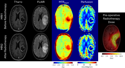 |
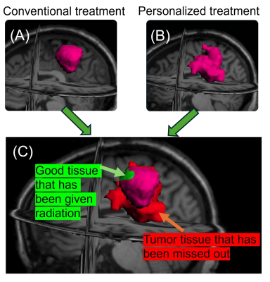
Computer Number: 142
2402. Dose
dependency of MRI changes following pre-operative radiotherapy
in glioblastoma; volumetric, ASL and APT changes
A. Fothergill, M. Waqar, A. Sovetkina, S. Parikh, D.
Higgins, O. Thomas, D. Coope, I. Djoukhadar, G. Borst, L.
Parkes
The University of Manchester, Manchester, United Kingdom
Impact: This study assesses early changes after
pre-operative brain irradiation in glioblastoma. We show
that hyperintense-FLAIR volume reduces, perfusion increases
and MTRasym decreases in a dose-dependent manner within 1-2
days of radiotherapy, providing potentially valuable
measures to track radiotherapy response.
|
|
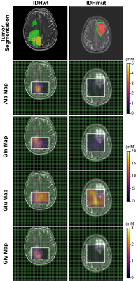 |
Computer Number: 143
2403. Distinct
Amino Acid Profiles in IDH Wild-Type and IDH-Mutant Glioma
Subregions: In Vivo 1H MRSI with Optimized Long-TE sLASER at 3T
S. Alcicek, M. W. Ronellenfitsch, J. P. Steinbach, K. J.
Weber, V. Prinz, M-T Forster, E. Hattingen, U. Pilatus, K.
J. Wenger
Goethe University, University Hospital Frankfurt, Frankfurt am Main, Germany
Impact: We used optimized long-TE-sLASER MRSI at 3T and
automated brain tumor segmentation to evaluate distinct
amino acid profiles in glioma molecular subtypes. Our study
highlights the necessity for separate (e.g.,
glutamate-glutamine, glycine-myoinositol) and
tumor-subregion-specific quantification of metabolites.
|
|
 |
Computer Number: 144
2404. Brain
Metabolic Alterations in Adult Survivors of Childhood Brain
Tumours Mapped through Multi-Voxel 1H MRS
S. Helsper, K. Bullens, C. Sleurs, A. Radwan, S. Sunaert, J.
Lemiere, S. Jacobs, U. Himmelreich
KU Leuven, Leuven, Belgium
Impact: Multi-voxel 1H
MR spectroscopy combined with corresponding anatomical MRI
can provide insight into metabolic brain alterations. Our
study suggests alterations related to axonal loss in adults
treated for childhood brain tumours, offering insights into
long-term effects of early-life cancer therapies.
|
 Back to Meeting Home
 Back to the Program-at-a-Glance
The International Society for Magnetic Resonance in Medicine is accredited by the Accreditation Council for Continuing Medical Education to provide continuing medical education for physicians.
 View
Presentation Video
View
Presentation Video