Digital Poster
Image Reconstruction Using AI
ISMRM & ISMRT Annual Meeting & Exhibition • 10-15 May 2025 • Honolulu, Hawai'i

 |
Computer Number: 17
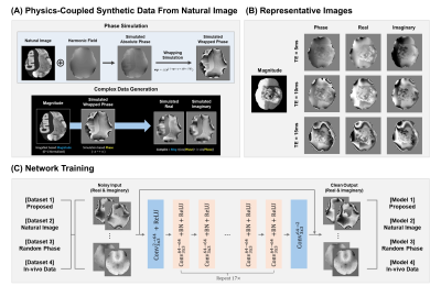
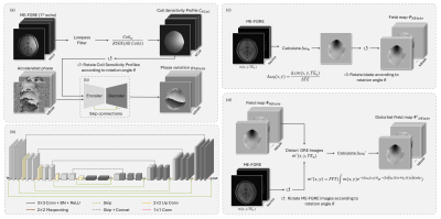
4602. Physics-Coupled
Synthetic Data Generation Method Using Natural Images Enabling
In-vivo Data-Free Complex MRI Denoising
S. Jung, D-H Kim
Yonsei University, Seoul, Korea, Republic of
Impact: Our method generates physics-coupled synthetic
data from natural images, enabling effective complex MRI
denoising without in-vivo data, achieving performance on par
with in-vivo-trained models. This approach reduces
dependence on large in-vivo datasets and addresses practical
challenges in in-vivo data collection.
|
|
 |
Computer Number: 18
4603. Single-breathhold
cardiac T1, T2, T2*, and fat fraction mapping at 0.55T using
dual rosette trajectory MRF and a deep image prior
reconstruction
E. Cummings, G. Lima da Cruz, J. Hamilton, N. Seiberlich
University of Michigan, Ann Arbor, United States
Impact: To reduce the time and number of breathholds
required for a cardiac scan, we introduce a method for
acquiring cardiac T1,
T2,
T2*,
and proton density fat fraction maps at 0.55T from a single
breathhold, 16-heartbeat sequence.
|
|
 |
Computer Number: 19
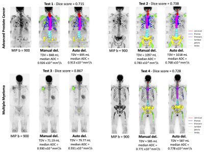
4604. Developing
a multi-channel deep-learning model for automatically
quantifying malignant bone disease from Multiparametric
Whole-Body MRI
A. Candito, R. Holbrey, L. D’Erme, S. Bottazzi, L. Russo, F.
Castagnoli, A. Dragan, C. Messiou, N. Tunariu, D-M Koh, M.
Blackledge
The Institute of Cancer Research, London, United Kingdom
Impact: Our multi-channel model can automatically
quantify TDV and ADC from suspected malignant bone lesions
across treatment with accuracy close to 70%, which can
assist with clinical decision-making in patients with
systemic cancer spread.
|
|
 |
Computer Number: 20
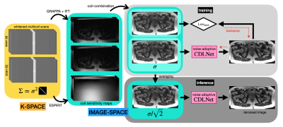
4605. Noise-Adaptive
MRI Denoising Using Self-Supervised Learning with
Average-to-Average (Avg2Avg) Loss
N. Janjusevic, M. Bruno, Y. Huang, J. Chen, Y. Wang, H.
Chandarana, L. Feng
Bernard and Irene Schwartz Center for Biomedical Imaging, New York University Grossman School of Medicine, New York, United States
Impact: The proposed denoising technique could greatly
encourage the use of 0.55T MRI and other low-SNR MRI
scanners, making imaging more affordable and accessible.
|
|
 |
Computer Number: 21
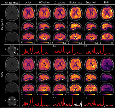
4606. Deep-ERx2:
Deep Learning Reconstruction for fast high-resolution
non-Cartesian Compressed-Sensing MR Spectroscopic Imaging at 3T
and 7T
P. Weiser, G. Langs, S. Motyka, B. Strasser, W. Bogner, P.
Golland, N. Singh, J. Dietrich, E. Uhlmann, T. Batchelor, D.
Cahill, G. Ungan, M. Hoffmann, A. Klauser, O. Andronesi
Athinoula A. Martinos Center for Biomedical Imaging, Massachusetts General Hospital, Boston, MA, USA, Cambridge, United States
Impact: Deep-ER enables high-resolution (3.4 mm
isotropic) metabolic imaging with clinically feasible
acquisition (4-9 min) and reconstruction times (1 min) at 3T
and 7T. These times are compatible with the clinical
workflow.
|
|
 |
Computer Number: 22
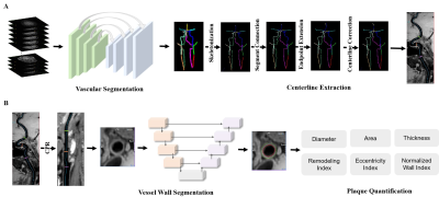
4607. Deep
learning for magnetic resonance vessel wall image: image
reconstruction, stenosis diagnosis and plaque calculation
f. fu, z. lin, x. yang, b. li
Ruijin Hospital affiliated to Shanghai Jiao Tong University School of Medicine, Shanghai, China
Impact: A deep learning algorithm for magnetic resonance
vessel wall interpretation accurately determined image
reconstruction, vessel stenosis and plaque calculation,
which achieved automatic postprocessing and had equivalent
diagnostic performance when compared with experienced
radiologists.
|
|
 |
Computer Number: 23
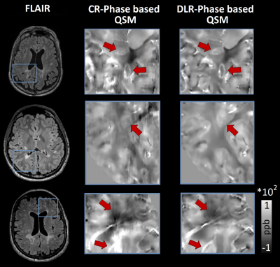
4608. Deep
Learning-reconstruction of rapid 3DEPI acquisitions for enhanced
QSM in the clinical assessment of Multiple Sclerosis
D. Gkotsoulias, M. Weigel, A. Cagol, N. de Oliveira Soares
Siebenborn, J. Pfeuffer, C. Granziera
University of Basel, Basel, Switzerland
Impact: Deep learning-based reconstruction, denoising
and super-resolution pipeline substantially enhances the
quality of QSM maps obtained from fast 3DEPI. This holds
promise for advancing the broader implementation of QSM in
the clinical management of Multiple Sclerosis.
|
|
 |
Computer Number: 24
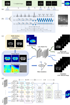
4609. High
efficient abdominal T1 and T2 mapping via LMC-MOLED acquisition
and GAL Multi-UNet reconstruction
Y. Zheng, W. Chen, Q. Lin, L. Zhu, L. Lin, J. Wang, Z. Chen,
S. Cai, C. Cai
Xiamen University, Xiamen, China
Impact: We proposed a new method for rapid simultaneous
T1 and
T2 mapping
of abdomen, addressing the issue of long acquisition time in
abdominal multi-parametric quantitative imaging, with
significant potential value for clinical diagnosis.
|
|
 |
Computer Number: 25
4610. Joint
attention deep learning reconstruction of highly-accelerated
pre- and post-contrast T1-weighted 3D images of brain tumors
A. Mekhanik, R. Otazo
Memorial Sloan Kettering Cancer Center, New York, United States
Impact: A
joint-attention deep learning reconstruction method can
exploit correlations across sequences and enable significant
reductions in MRI protocols.
|
|
 |
Computer Number: 26
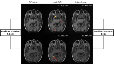
4611. Fast
and accurate reconstruction of accelerated 7T
susceptibility-weighted imaging using multi-scale hybrid
CNN-Transformer network
C. Duan, D. Zhang, X. Bian, J. Qu, X. Lou
The First Medical Center, Chinese PLA General Hospital, Beijing, China
Impact: The potentially reduced scan time with MSCT-Net
offers new possibilities for widely adoption of 7T SWI in
clinical brain imaging. Furthermore, the approach has the
potential to guide design of reconstruction models for other
high-resolution 7T MRI data.
|
|
 |
Computer Number: 27
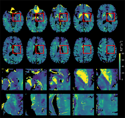
4612. Accelerated
T2* Mapping of the Human Brain at 7T Using Deep Learning:
Achieving 0.6 mm Isotropic Resolution in Under 6 Minutes
A. Klauser, E. Sleight, T. Yu, N. Pato Montemayor, J.
Phillippe, D. Nickel, L. Bacha, T. Di Noto, B. Maréchal, T.
Kober, T. Hilbert, G. F. Piredda
Siemens Healthineers, Lausanne, Switzerland
Impact: We demonstrate the efficacy of deep
learning-based reconstruction for highly accelerated
acquisitions, enabling 0.6mm isotropic R2* mapping of the
brain in 6 minutes at 7T. This method highlights
submillimeter T2* contrast, potentially enhancing its
application in detecting microstructural alterations.
|
|
 |
Computer Number: 28
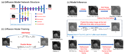
4613. CRNN
with Bidirectional Frame-By-Frame Diffusion-Model-Based
Refinement for Cardiac cine MRI Reconstruction
H. Li, H. Sun, Z. Li, R. Yang, H. Chen
Tsinghua University, Beijing, China
Impact: A novel diffusion-model-based method of
accelerated cardiac cine MRI reconstruction has been
developed and improved the quality of reconstructed images.
This may facilitate the application of accelerated cardiac
cine MRI in clinical medicine, reducing patient discomfort
and motion related artifacts.
|
|
 |
Computer Number: 29
4614. AI-assisted
Collaborative Reconstruction for Highly-accelerated
DW-PROPELLER-EPI
H. Xiong, L. Liang, S. Chen, X. Xu, C. Yuan, Y. Li, T. Liu,
H-C Chang
The Chinese University of Hong Kong, Hong Kong, China
Impact: High geometric fidelity and high-resolution
brain DWI with reasonable scan time may benefit clinical
applications and neuroscience research.
|
|
 |
Computer Number: 30
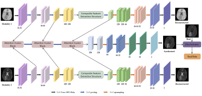
4615. Robust
Image Synthesis Method of Multi-Modal MRI Utilizing a
Transformer Architecture
H. Zhang, H. Guo, M. Li, X. Liu
Shenyang University of Technology, Shenyang, China
Impact: This study introduces a novel technique for
generating missing modality high quality images in MRI,
which is robust to motion artifacts. The provision of
structurally intact images enables clinicians to identify
lesions more efficiently, augment diagnostic precision.
|
|
 |
Computer Number: 31
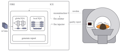
4616. Integrating
Inline Quality Control at the MRI Scanner: Global and Local
Assessment of Motion Artifacts Using Deep Learning
V. Ecker, M. Ganz, H. Eichhorn, E. Marchetto, T. Huelnhagen,
B. Yang, S. Gatidis, T. Küstner
University Hospital of Tübingen, Tübingen, Germany
Impact: Our inline integration assessment for global and
local image quality in MRI scans enables reliable detection
of motion artifacts. This advancement allows for immediate
corrective actions, improving diagnostic accuracy and
optimizing imaging workflows.
|
|
 |
Computer Number: 32
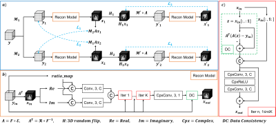
4617. Unsupervised
reconstruction of highly undersampled 3D cones cardiac image
navigators using a dual-branch joint training framework
X. Guo, C. Sheagren, J. Patel, L. Li, G. Wright, F. Guo
Huazhong University of Science and Technology, Wuhan, HuBei, China
Impact: Our approach provides
a way
to reconstruct highly undersampled 3D
cardiac images with sufficient quality for
retrospective motion correction, suggesting the utility of
our approach in various scenarios where fully sampled data
is unavailable.
|
 Back to Meeting Home
 Back to the Program-at-a-Glance
The International Society for Magnetic Resonance in Medicine is accredited by the Accreditation Council for Continuing Medical Education to provide continuing medical education for physicians.
 View
Presentation Video
View
Presentation Video