Digital Poster
Imaging Biomarkers in MS: Diagnosis & Monitoring
ISMRM & ISMRT Annual Meeting & Exhibition • 10-15 May 2025 • Honolulu, Hawai'i

Digital Poster
Imaging Biomarkers in MS: Diagnosis & Monitoring
 |
Computer Number: 81
3579. Longitudinal
Analysis of [11C]DPA-713 PET and QSM in Multiple Sclerosis for
Differentiating the Spatial Patterns of Pathological Lesions
Y. Lee, Y. Kang, S. Hurtado Rua, T. Nguyen, S. Gauthier
Pusan National University, Busan, Korea, Republic of
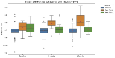
Impact: Our analysis suggests that the early-stage
inflammatory activity in the lesion center is associated
with the development of a paramagnetic rim.
|
|
 |
Computer Number: 82
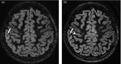
3580. Myelin
dynamics in acute multiple sclerosis lesions: Short-term myelin
water changes relate to long-term lesion outcome
I. Vavasour, A. Traboulsee, D. Li, S. Kolind, A. Rauscher,
G. W. Moore, A. MacKay, C. Laule
University of British Columbia, Vancouver, Canada
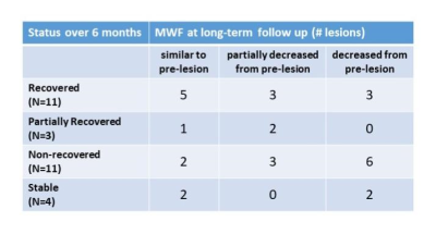
Impact: Early myelin water fraction dynamics in new
multiple sclerosis may predict lesion MWF at 5 years.
|
|
 |
Computer Number: 83
3581. Automatic
Segmentation of Diffusely Abnormal White Matter in Multiple
Sclerosis using Fuzzy C-means Clustering
T. Joseph, S. Balaji, S. Kolind, G. Zhao, P. Sun, R.
Carruthers, A. Schabas, A-L Sayao, V. Devonshire, R. Tam, G.
R. W. Moore, D. K. B. Li, A. Traboulsee, I. Vavasour, C.
Laule
University of British Columbia, Vancouver, Canada
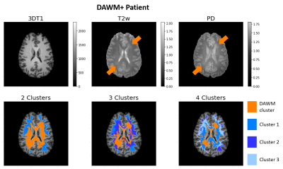
Impact: Unsupervised clustering could provide an
unbiased approach to automatically segment diffusely
abnormal white matter (DAWM) in multiple sclerosis patients
in vivo. In this study, the subtle hyperintensities
associated with DAWM were segmented on proton density and T2-weighted
images.
|
|
 |
Computer Number: 84
3582. Choroid
Plexus Radiomics for Differentiating MS and NMOSD and Their
Correlation with Clinical Characteristics
X. Wang, X. Wang, Y. Liu, Y. Li
The First Affiliated Hospital of Chongqing Medical University; Chongqing Medical University, Chongqing, China
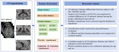
Impact: This research advances the understanding of CP
abnormalities in MS and NMOSD, aiding differential diagnosis
and clinical monitoring.
|
|
 |
Computer Number: 85
3583. Integrating
BMAT and FA Imaging for Improved Classification of Multiple
Sclerosis: A Machine Learning Perspective
C. Montalba, P. Franco, R. Caulier-Cisterna, M. Vasquez, C.
Cárcamo, E. Ciampi, M. Andia
Pontificia Universidad Católica de Chile, Santiago, Chile., Santiago, Chile
Impact: This study enhances diagnostic precision for MS,
facilitating early intervention and personalized treatment
strategies. It encourages further exploration of cognitive
decline mechanisms in MS and may inspire similar integrative
approaches in other neurological disorders, ultimately
improving patient outcomes.
|
|
 |
Computer Number: 86
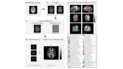
3584. Retrospective
Analysis of Diffusion Tensor Imaging from the SPRINT-MS Clinical
Trial: Advancing Trial Methodology
K. Sakaie, M. Du, M. Lowe, J. Lin, R. Fox
The Cleveland Clinic, Cleveland, United States
Impact: The cingulum and cerebellar peduncle are
potential areas for focus in the evaluation of therapies in
progressive MS.
|
|
 |
Computer Number: 87
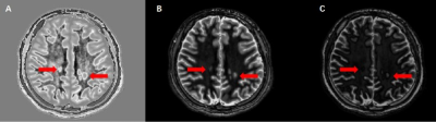
3585. Enhanced
Evaluation of Juxtacortical Lesions in Multiple Sclerosis Using
High-Resolution Double Inversion Recovery Imaging with Deep
Learning
T. Shintaku, S. Ide, H. Nagaya, Y. Ishimoto, K. Watanabe, K.
Oyu, S. Kasai, Y. Umemura, M. Sasaki, K. Saitou, A. Ozawa,
A. Nozaki, X. Zhu, T. Wakayama, H. Nishijima, C. Suzuki, M.
Tomiyama, S. Kakeda
Hirosaki University Graduate School of Medicine, Hirosaki, Aomori, Japan
Impact: 3D DL-Speed double inversion recovery imaging is
acquired in 4:23 with 0.7mm isotropic resolution covering
the whole brain, which could improve the accuracy of
cortical lesion assessment in patients with multiple
sclerosis.
|
|
|
Computer Number:
3586. WITHDRAWN |
||
 |
Computer Number: 88
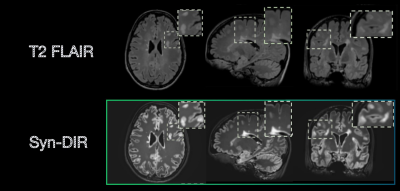
3587. Synthesis
of Double Inversion Recovery-like Images for Superior Contrast
and Multiple Sclerosis Lesion Visibility
L. Wang, C. Arnold, Z. Zhou, L. Xiang, A. Shankaranarayanan,
S. Bash, L. Tanenbaum
Subtle Medical, Menlo Park, United States
Impact: Syn-DIR provides superior lesion visibility and
consistent brain structure representation. The strong
agreement in quantitative lesion and regional brain volumes
demonstrates its robustness for clinical and research
applications.
|
|
 |
Computer Number: 89
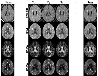
3588. Susceptibility
Source Separation for Quantification of Remyelination in
Multiple Sclerosis Lesions from multi Gradient-Echo (mGRE) Data
M. Sisman, A. Wu, H. Luu, A. Dimov, H. Schwartz, K.
Markowitz, I. Pliska-Bloch, P. Spincemaille, Y. Wang, S.
Gauthier, T. Nguyen
Cornell University, New York, United States
Impact: Understanding about the mechanism and timing of
the remyelination in multiple sclerosis (MS) is incomplete.
Therefore, noninvasive quantification of myelin is fast and
accurately is an important clinical need. Susceptibility
source separation from gradient-echo carries potential to
satisfy this need.
|
|
 |
Computer Number: 90
3589. Assessment
of multiple sclerosis using divided subtracted inversion
recovery (dSIR) technique: a quantitative study
J. Huang, J. Wang, Y. Shan, C. Zhao, B. Xu, Y. Ma, J. Lu
Xuanwu hospital, Beijing, China
Impact: The dSIR sequence can effectively quantitatively
evaluate the abnormal changes of deep GM nuclei and WM
lesions in MS patients and may be used for clinical
monitoring of patients' cognitive and disability status.
|
|
 |
Computer Number: 91
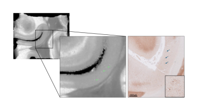
3590. 9.4T
MRI-derived Myelin Water Fraction in post-mortem brain supports
the identification of active remyelination in Multiple Sclerosis
I. Callegari, D. Gkotsoulias, J. Leupold, E. Bahn, J. Franz,
B. Dhital, D. von Elverfeldt, V. Kiselev, M. Weigel, C.
Stadelmann, C. Granziera
Department of Biomedical Engineering, Basel, Switzerland
Impact: An unprecedented MWF with ultra-high spatial
resolution obtained in post-mortem brain blocks imaged 9.4T
MRI enabled the detection of ongoing remyelination at an
almost cellular level, opening a new window into repair
mechanisms in MS.
|
|
 |
Computer Number: 92
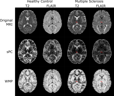
3591. Characteristics
of MRI Phase Congruency Texture in Limbic Brain Regions relates
to Depression in People with Comorbid MS
O. Oladosu, Y. Zhang
University of Calgary, Calgary, Canada
Impact: Non-invasive measurement of subtle structural
changes in depression-relevant brain gray matter may be
invaluable for improving our understanding of the underlying
mechanisms in people with comorbid MS and depression,
encouraging further investigations of MRI phase congruency
analyses.
|
|
 |
Computer Number: 93
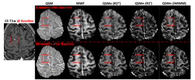
3592. QSM
Source Separation Detects Treatment Effect of Ocrelizumab on
Multiple Sclerosis Lesion Myelin: A Longitudinal Study
M. Sisman, K. Markowitz, H. Luu, H. Schwartz, I.
Pliska-Bloch, P. Spincemaille, I. Kovanlikaya, S. Hurtado
Rua, S. Gauthier, Y. Wang, T. Nguyen
Cornell University, New York, United States
Impact: Treatment efforts in Multiple Sclerosis includes
medications that suppress acute inflammation. Therefore, the
noninvasive quantification of the effect of treatment on the
iron change and remyelination carries significant
importance. Here, a novel gradient-echo based approach is
tested for this purpose.
|
|
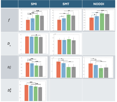 |
Computer Number: 94
3593. Comparative
Evaluation of Microstructural Diffusion Methods in
Characterizing Multiple Sclerosis Lesions
C. Jin, A. Toubasi, C. Gheen, T. Vinersky, X. Jiang, F.
Bagnato, J. Xu
Vanderbilt University Institute of Imaging Science, Nashville, United States
Impact: This study provides a better understanding of
how multiple widely-used diffusion methods characterize MS
lesions, which can assist in identifying the most effective
imaging techniques for more accurate diagnoses in MS
clinical practice.
|
|
 |
Computer Number: 95
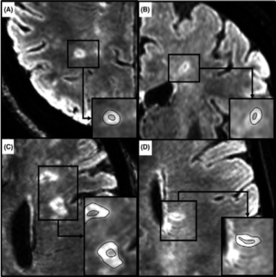
3594. Characterizing
Lesion Activity in Multiple Sclerosis through Central Vein Sign
and Diffusion Tensor Imaging
S. Hannoun, G. Fayad, N. Al-Arab, F. Cotton
American University of Beirut, Beirut, Lebanon
Impact: CVS presence in MS lesions significantly aids in
distinguishing active, chronic lesions, enabling more
precise monitoring of disease progression and management.
This study highlights CVS’s diagnostic value, warranting
routine MRI inclusion in MS protocols.
|
 Back to Meeting Home
 Back to the Program-at-a-Glance
The International Society for Magnetic Resonance in Medicine is accredited by the Accreditation Council for Continuing Medical Education to provide continuing medical education for physicians.
 View
Presentation Video
View
Presentation Video