Digital Poster
Mapping Brain Changes in Multiple Sclerosis
ISMRM & ISMRT Annual Meeting & Exhibition • 10-15 May 2025 • Honolulu, Hawai'i

Digital Poster
Mapping Brain Changes in Multiple Sclerosis
 |
Computer Number: 97
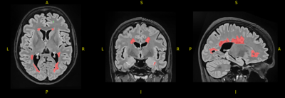
3753. Glymphatic
Alterations Mediate Cognitive Dysfunction in Relapsing-Remitting
Multiple Sclerosis
Z. Wang, X. Xu, W. Ren, J. Liu, J. Zhang, K. Ai
The Second Hospital & Clinical Medical School, Lanzhou University, Lanzhou, China
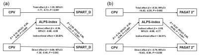
Impact: The impact of enlarged CP on cognitive function
is mediated by glymphatic alterations, indicating a
pathological mechanism for MS-related cognitive impairment.
|
|
 |
Computer Number: 98
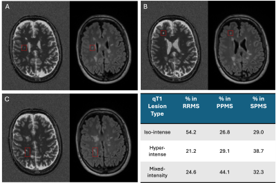
3754. A
quantitative MRI-Based Normative Framework for Personalizing
Microstructural Pathology in Multiple Sclerosis
X. Chen, P-J Lu, M. Ocampo-Pineda, A. Cagol, M. Weigel, S.
Schädelin, K-S Chan, M. Zwiers, J. Kuhle, L. Kappos, L.
Melie-Garcia, J. Marques, C. Granziera
Translational Imaging in Neurology (ThINK) Basel, Department of Biomedical Engineering, Faculty of Medicine, University Hospital Basel and University of Basel, Basel, Switzerland
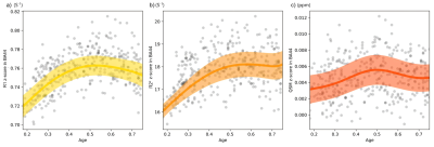
Impact: This qMRI-based normative model enables
individualized quantification of multiple sclerosis
pathology, which is strongly related to clinical measures
and a fluid biomarker of neuroaxonal damage, opening a new
perspective for clinical stratification and personalized
treatment decisions.
|
|
 |
Computer Number: 99
3755. Quantifying
longitudinal tract profile differences in multiple sclerosis
lesional tracts
A. Wu, A. Traboulsee, D. Li, R. Tam, J. Oh, I. Vavasour, P.
Johnson, N. Wiley, E. Gallinger, A. MacKay, S. Kolind, S.
Balaji
University of British Columbia, Vancouver, Canada
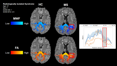
Impact: Differences in lesional tract profiles of myelin
water fraction and fractional anisotropy were quantified
across two timepoints twelve months apart using two newly
proposed measures in people with multiple sclerosis, and
showed moderate correlations with clinical scores.
|
|
|
Computer Number:
3756. WITHDRAWN |
||
 |
Computer Number: 100
3757. Ultra-High
Contrast (UHC) MRI of the Brain and Spinal Cord in Multiple
Sclerosis Using Divided Subtracted Inversion Recovery Sequences
P. Condron, D. Cornfeld, M. Bydder, C. Shi, T. Emsden, K.
Whitehead, G. Newburn, M. Scadeng, S. Holdsworth, G. Bydder
Mātai Medical Research Institute , Gisborne, New Zealand
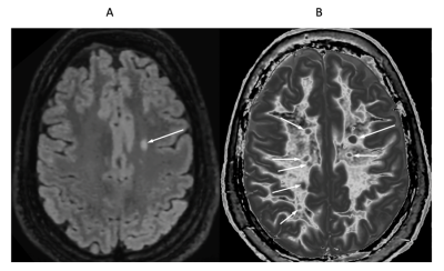
Impact: Ultra-high contrast (UHC) divided subtracted
inversion recovery (dSIR) sequences were used in patients
with multiple sclerosis during relapse and in remission.
Well defined focal lesions and very widespread abnormalities
in white matter were shown when conventional imaging
appeared normal.
|
|
 |
Computer Number: 101
3758. Correlation
between lesion volume and total brain, thalami and caudate
volumes obtained by automatic volumetry in multiple sclerosis
patients.
F. Ayala-Ochoa, A. Hernandez-Medina, B. Elias-Perez, E.
Torres-Olivas
Hospital Angeles Lomas, Huixquilucan, Mexico
Impact: Total lesion burden in MS patients correlates
with structural brain volume loss mediated by destruction of
white matter tracts and Wallerian degeneration. Establishing
volumetric biomarkers using artificial intelligence may
provide useful information regarding disease progression and
long-term prognosis.
|
|
 |
Computer Number: 102
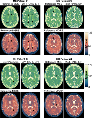
3759. Simultaneous
T2 and T2* Mapping in Multiple Sclerosis using Accelerated
2in1-RARE-EPI and Model-Based Reconstruction
J. R. Velasquez Vides, T. Gladytz, J. Millward, S. Waiczies,
S. Shalikar, J. Kuchling, F. Paul, L. Krenz, G. Rose, T.
Niendorf
Max-Delbrück-Center for Molecular Medicine in the Helmholtz Association, Berlin, Germany
Impact: 2in1-RARE-EPI combined with model-based
reconstruction methods provides a technical foundation
supporting clinical qMRI of neuroinflammatory and
neurodegenerative diseases. It also has the potential for
accelerated and simultaneous T2 and
T2*
mapping of the myocardium, eyes, and kidneys.
|
|
 |
Computer Number: 103
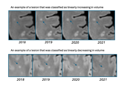
3760. Patterns
of Lesion Volume Evolution in Multiple Sclerosis: Associations
with Microstructural Change and Neuroaxonal Damage
M. Greselin, P-J Lu, A. Cagol, E. Ruberte, M. Pineda, S.
Schaedelin, L. Melie-Garcia, X. Chen, R. Galbusera, M.
Weigel, J. Kuhle, L. Kappos, H. Ganjgahi, C. Granziera
University Hospital Basel and University of Basel, Basel , Switzerland
Impact: These findings can reflect the importance of
chronic neurodegenerative processes within MS lesions and
provide a foundation for further investigation into the
mechanisms of disease progression and clinical worsening in
pwMS.
|
|
 |
Computer Number: 104
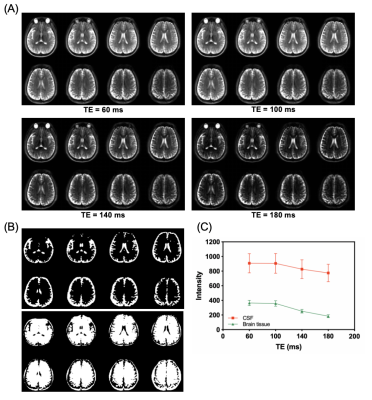
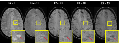
3761. Potential
application of magnetization transfer-indirect spin labeling MRI
for imaging multiple sclerosis at 3T
Z. Wang, P. Cai, J. Wang, H. Zhang, S. Zeng, J. Huang
The University of Hong Kong, Hong Kong, China
Impact: This study optimized the echo time for MISL MRI
of human brain at clinical 3T. MISL MRI detected a
decreasing trend in water exchange between CSF and brain
tissue in MS patients, suggesting its potential for imaging
neurological diseases.
|
|
 |
Computer Number: 105
3762. Quantitative
T1 as a Marker of Myelin Content in Multiple Sclerosis: A
Genome-Wide Association Study
N. Bar Zohar, A. Cagol, M. Ocampo-Pineda, D. Gkotsoulias,
P-J Lu, T. Sirito, D. Gindes, X. Chen, M. Weigel, S. Cichon,
J. Kuhle, L. Kappos, C. Granziera
University Hospital and University of Basel, Basel, Switzerland
Impact: This study provides insight into genetic
influences on remyelination in MS, highlighting loci that
may guide personalized therapeutic approaches. These
findings could lead to new research into genetic predictors
of repair capacity, potentially advancing targeted
treatments for neuroprotection and recovery.
|
|
 |
Computer Number: 106
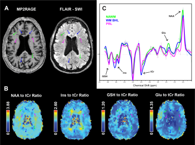
3763. 7T
3D-MR Spectroscopic Imaging of GSH and GPX4 Immunohistochemistry
Staining Reveal Oxidative Stress Patterns in MS Lesions

R. Rumbak, A. Dal-Bianco, F. Niess, B. Strasser, L.
Hingerl, A. Kloss-Brandstätter, S. Hametner, R.
Höftberger, T. Berger, G. Grabner, P. Rommer, W. Bogner,
E. Niess
Medical University of Vienna, Vienna, Austria
Impact: High-resolution 7T 3D-MRSI mapping of GSH
reveals oxidative stress patterns across MS lesions,
enhancing in vivo lesion
monitoring and offering comprehensive insights that support
clinical management and progression tracking in multiple
sclerosis.
|
|
 |
Computer Number: 107
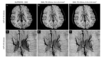
3764. SUPREME:
Susceptibility with Ultra-high-resolution Parallel
REconstruction of Multi-Echo
V. Truong, H. Sawan, S. Wright, Y. Chen
Wayne State University School of Medicine, Detroit, United States
Impact: This study demonstrates the imaging capabilities
of SUPREME to quantify medullary vein density and venous
oxygen saturation, and central vein lesion load which will
be used in multiple sclerosis studies.
|
|
 |
Computer Number: 108
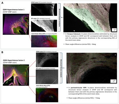
3765. Imaging
properties of astrocytic activation in Multiple Sclerosis using
MRI and structural tensors

D. Gkotsoulias, I. Callegari, P-J Lu, E. Bahn, J. Franz,
M. Weigel, C. Stadelmann, L. Kappos, C. Granziera
University of Basel, Basel, Switzerland
Impact: We provide initial evidence that QSM
contrast might also be sensitive to astrocytic activation in
MS lesions and surrounding NAWM. The paternalistic
activation of astrocytes which resembles local WM axonal
directionality introduces a completely novel
viewpoint in MS disease progression.
|
|
 |
Computer Number: 109
3766. Optimized
Susceptibility Weighted Imaging for the Detection of Central
Vein Sign in Multiple Sclerosis at 3-tesla
S. Madhusoodhanan Nair, Y-C Hsu, E. Luskin, D. Ayrapetyan,
A. Liu, N. Binesh, M. Maya, Y. Xie, D. Li, M. Kaisey, O.
Al-Louzi, D. Ontaneda, N. Sicotte, P. Sati
Cedars-Sinai Medical Center, Los Angeles, United States
Impact: Optimized SWI protocol increases CVS detection
and improves diagnostic classification for MS as compared to
standard SWI.
|
|
 |
Computer Number: 110
3767. On
the Impact of QSM Pipeline Parameters on Brain Iron Outcomes
F. Salman, N. Bergsland, M. Dwyer, B. Weinstock-Guttman, R.
Zivadinov, F. Schweser
Buffalo Neuroimaging Analysis Center, Department of Neurology at the Jacobs School of Medicine and Biomedical Sciences, University at Buffalo, The State University of New York, Buffalo, United States
Impact: Our findings suggest that neither younger age
nor specific regularization parameter choices account for
the discrepancies in the literature, and that QSM robustly
detects disease-related effects independent of these
parameter variations.
|
|
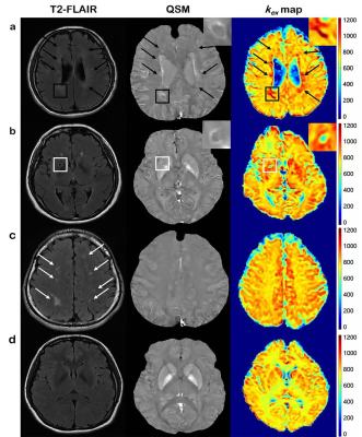 |
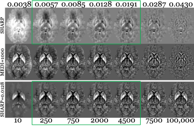
Computer Number: 111
3768. Quantify
oxidative stress and iron via kex MRI and QSM for differential
diagnosis of multiple sclerosis and cerebral small vessel
diseases.
M. Hou, H. Li, Y. Jiao, W. Chen
Tongji Hospital of Tongji Medical College of Huazhong University of Science and Technology, Wuhan, China
Impact: The in vivo visualization and quantification of
oxidative stress and iron deposition via kex MRI
and QSM are the potential biomarkers to promote the early
differential diagnosis of MS and SVD.
|
 Back to Meeting Home
 Back to the Program-at-a-Glance
The International Society for Magnetic Resonance in Medicine is accredited by the Accreditation Council for Continuing Medical Education to provide continuing medical education for physicians.
 View
Presentation Video
View
Presentation Video