Digital Poster
Data Acquisition
ISMRM & ISMRT Annual Meeting & Exhibition • 10-15 May 2025 • Honolulu, Hawai'i

 |
Computer Number: 1
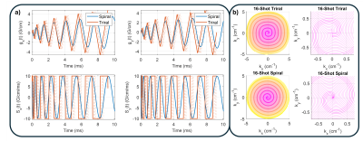
3184. Trirals
for Imaging 10% Faster
M. McCready, D. Abraham, Z. Shah, K. Setsompop, J. Pauly, A.
Kerr
Stanford University, Stanford, United States
Impact: Decreasing the readout time using trirals is
important for time sensitive artifacts, such as field
imperfections, and sequence TR/TE dependent signal changes.
|
|
 |
Computer Number: 2
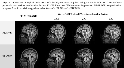
3185. Evaluation
of Wave-CAIPI for accelerating MP2RAGE and FLAWS in deep-brain
nucleus localization
C. Liu, S. Wang, Y. Wu, D. N. Splitthoff, W-C Lo, P. Hang,
J. Sun
Department of Radiology, Second Affiliated Hospital of Zhejiang University, School of Medicine, Hangzhou, China
Impact: The Wave-CAIPI 2x2 and 2x3 protocols markedly
enhanced MRI efficiency, improving patient comfort and
accessibility while maintaining high-resolution imaging
essential for accurate deep-brain structure visualization in
clinical practice.
|
|
 |
Computer Number: 3
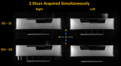
3186. Design
of a differential single-band presaturated ultrashort echo time
(dSB-UTE) sequence for accelerated short-T2 imaging
J. Reich, K. Harkins, R. Crescenzi, E. MacMillan, R. Feldman
University of British Columbia, Kelowna, Canada
Impact:
The dSB-UTE sequence is expected to reduce scan times for short-T2 imaging by a factor of 1.55-4.72 while achieving the same echo times as current UTE sequences. The dSB-UTE shows improved image quality over other simultaneous multi-slice UTE techniques. |
|
 |
Computer Number: 4
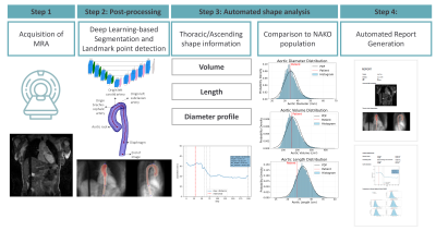
3187. Clinical
Translation of Deep Learning-based Ascending Aortic Morphology
Characterization Using 3D Non-Contrast-enhanced MRA
L. Fay, D. Amsel, V. Ecker, M. Lescan, T. Hülnhagen, D.
Giese, B. Yang, S. Gatidis, T. Kuestner
Medical Image and Data Analysis (MIDAS.lab), Department of Diagnostic and Interventional Radiology, University Hospital of Tuebingen, Tuebingen, Germany
Impact: Integration of deep learning image analysis for
thoracic aorta inline on the MR scanner accelerates aortic
morphology assessment across multiple sequences. Visualizing
the results directly on the scanner supports rapid clinical
decisions and advances cardiovascular imaging workflows.
|
|
 |
Computer Number: 5
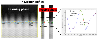
3188. Pilot
Tone-based Diaphragm Motion Estimation for Navigator-Triggered
Magnetic Resonance Cholangiopancreatography
X. Li, B. Kühn, W. Majeed
Siemens Medical Solutions USA, Inc., Saint Louis, United States
Impact: This study demonstrates that the pilot
tone-based diaphragm motion estimation framework enables
estimation of motion information associated with
navigator-triggered MRCP acquisitions. These results have
potential to improve image quality and workflow of MRCP
scans.
|
|
 |
Computer Number: 6
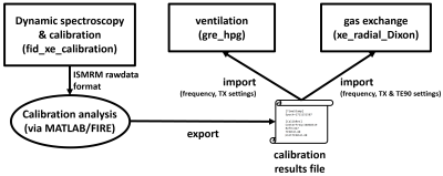
3189. Improved
Workflow for Hyperpolarized 129Xe MRI through Automated
Adjustments
J. Mugler III, L. Cui, K. Chow, A. Costelle, S.
Leewiwatwong, J. Mata, Y. Shim, P. Niedbalski, B. Driehuys,
R. Eddy
University of Virginia School of Medicine, Charlottesville, United States
Impact: The
proposed automated adjustment-processing framework results
in a workflow for xenon lung MRI that mirrors that for
routine clinical proton MRI, increasing the accessibility of
hyperpolarized 129Xe
lung MRI and facilitating its translation into clinical use.
|
|
 |
Computer Number: 7
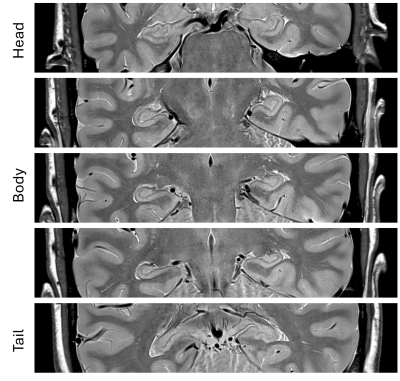
3190. Improving
Hippocampus Volumetry Using a Deep Learning-based Proton
Density-Weighted TSE sequence at 3T
S. Buch, V. Truong, Y. Xuan, R. Gattu, Y. Chen
Wayne State University, Detroit, United States
Impact: This study demonstrates that the deep learning
based 2D high-resolution proton density weighted TSE
sequence has a potential to reduce inaccuracies in
hippocampus volumetry, which will ensure reliable diagnosis
and monitoring of neurological and psychiatric conditions.
|
|
 |
Computer Number: 8
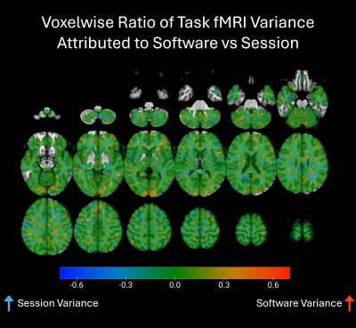
3191. Test-retest
reliability of magnetic resonance imaging data across the
Siemens VE11C to XA30 software upgrade
K. Jones, S. Noble, J. Gilbert, M. Yen, R. Welsh
University of California, Los Angeles, Los Angeles, United States
Impact: We found no meaningful difference between MRI
datasets collected using the Siemens VE11C and XA30 software
packages. This evidence of consistency in longitudinal MRI
data across software lends validity to imaging studies that
are affected by unforeseen software upgrades.
|
|
 |
Computer Number: 9
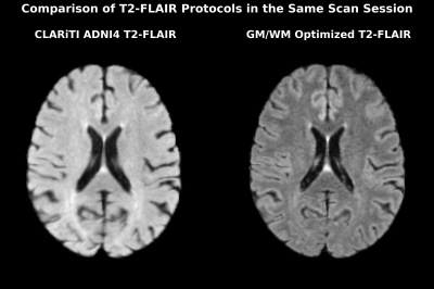
3192. Inter-Scanner
Variability and Evaluation of T2-FLAIR Harmonization in
Alzheimer’s Neuroimaging
B. Ho, D. Kim, A. Kumar, S. Weiss, H. Vossler, A. Dreisbach,
D. Holley, M. Khalighi, E. Mormino, G. Zaharchuk
Stanford University, Stanford, United States
Impact: T2-FLAIR is an instrumental imaging sequence to
visualize gray-white matter contrast and white matter
abnormalities. However, inter-scanner variability may hinder
analysis involving multi-site imaging datasets. Thus, we
evaluated protocol changes that can improve harmonization of
T2-FLAIR images across scanners.
|
|
 |
Computer Number: 10
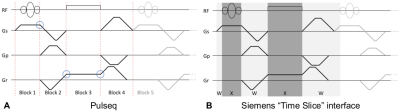
3193. Python-based
Pulseq Client to the Remote Sequence Streaming Interface
M. Shafiekhani, T. Kluge, B. Wilhelm-Feldbusch, P. Hucker,
C. Forman, R. Schneider, M. Zaitsev
University Medical Center Freiburg, Freiburg, Germany
Impact: Interpreter modules are instrumental for
executing open pulse sequences on a particular scanner. The
presented work paves a way towards reducing the effort
associated with their development and maintenance by
potentially reducing adaptations and tests for every scanner
software version.
|
|
 |
Computer Number: 11
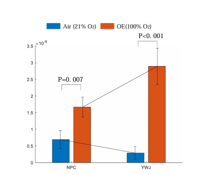
3194. Clinical
Feasibility and Tolerance of Oxygen-Enhanced MRI for Assessing
Tumor Hypoxia in Nasopharyngeal Carcinoma
F. Chen, J. Li, S. x. Xiang, W. Chen
Department of Radiology, Southwest Hospital, Army Medical University , Chong Qing, China
Impact: Dynamic T1-mapping with OE-MRI to indicate
hypoxia of NPC and the derived parameter ΔR1 may be a
repeatable hypoxia biomarker. OE-MRI is promising to guide
clinical work of biology guided adaptive radiotherapy.
|
|
 |
Computer Number: 12
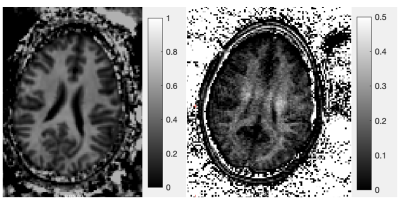
3195. Towards
ihMT Imaging at 7T Using 3D Centric Gradient Echo Readout for
ihMT Contrast Optimization: Preliminary Results
P. Will, W. Bogner, E. Niess, F. Niess, M. Zaiss, N.
Weiskopf, T. Emmenegger
Department of Biomedical Imaging and Image-Guided Therapy, Highfield MR Center, Medical University of Vienna, Vienna, Austria, Vienna, Austria
Impact: Acquiring 7T ihMT with a 3D centric spiral
gradient echo readout potentially enhances specificity and
contrast between WM and GM. This approach has the potential
to improve the myelin contrast, enabling more precise
clinical assessments in spinal neurological pathologies.
|
|
 |
Computer Number: 13
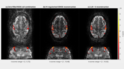
3196. Sub
2 mm resolution fMRI at 3T using randomly undersampled 3D-EPI
with locally low-rank + temporally sparse reconstruction
R. Fung, R. Lobos, J. Fessler, D. Noll, J-F Nielsen
University of Michigan, Ann Arbor, United States
Impact: Randomized 3D-EPI and locally low-rank plus
temporally sparse decomposition are novel approaches for
high resolution fMRI. Other fMRI scientists can use our
vendor-agnostic, open-source implementation as a template,
adapting it to suit their specific high resolution fMRI
needs.
|
|
 |
Computer Number: 14
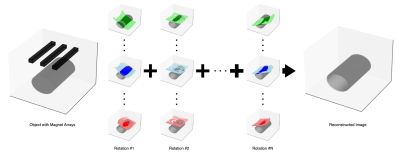
3197. Rotating
Magnet Arrays as Spatial Encodings for Portable MRI Applications
Y. Shi, N. Ayar, K-J Jung, K. Haran
University of Illinois Urbana-Champaign, Champaign, United States
Impact: The preliminary prototype embraces inhomogeneity
in the base magnetic field, eliciting a new direction for
MRI instrumentation. Its ability to produce pilot scans
allows for early diagnosis while reducing costs and
providing patient comfort, ultimately making MRI technology
more accessible.
|
|
 |
Computer Number: 15
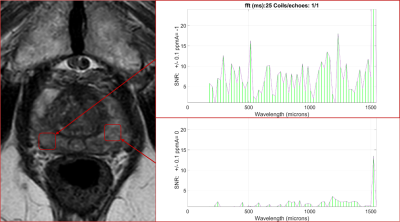
3198. Magnetic
Resonance Histopathology (MRH): a direct, noninvasive, and
low-cost diagnostic for quantitative microarchitecture imaging.
T. James, A. Benjamin, S. Ma, R. Garipov, G. Crelier, Q. Lu,
F. Han, D. Turley, K. James, N. Sta Maria, K. Williams, I.
Parker, R. Jacobs
BioProtonics,Inc., Santa Barbara, United States
Impact: MRH provides a currently inaccessible
quantitative measure of sentinel tissue microscopic
characteristics enabling more informed diagnosis, reducing
the need for excisional observation.
|
|
 |
Computer Number: 16
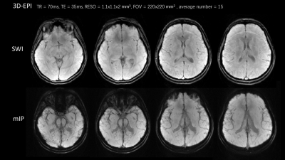
3199. 3D-EPI
SWI at 0.5T
Y. Gao, Y. Lian, Y. Jiang, J. Liu, Z. Huang, Y. Ye, H. Guo
Center for Biomedical Imaging Research, School of Biomedical Engineering, Tsinghua University, Beijing, China
Impact: 0.5T with 3D-EP-SWI offers greater access than
higher field strength MRI, and may meet clinical needs for
diagnosis of cerebral diseases like cerebral microbleeds.
|
 Back to Meeting Home
 Back to the Program-at-a-Glance
The International Society for Magnetic Resonance in Medicine is accredited by the Accreditation Council for Continuing Medical Education to provide continuing medical education for physicians.
 View
Presentation Video
View
Presentation Video