Digital Poster
Spinal Cord
ISMRM & ISMRT Annual Meeting & Exhibition • 10-15 May 2025 • Honolulu, Hawai'i

 |
Computer Number: 81
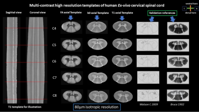
1610. High-Resolution
Multi-Contrast Template Construction of the Cervical Spinal Cord
Using Anatomical and Diffusion MRI at 80 µm
I. Hattan, G. Cowin, N. Kurniawan
Ministry of Health (MOH) and Ministry of Education (MOE), Jazan, Saudi Arabia
Impact: This high-resolution multi-contrast template can
potentially improve anatomical and microstructural analysis
disease-related changes. For example, besides measuring
atrophies, clinicians could use the template to pin-point
subtle changes in the GM motoneuron pool due to degeneration
or injuries.
|
|
 |
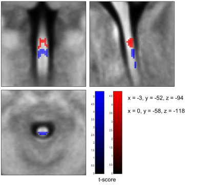
Computer Number: 82
1611. Rootlets-informed
registration to a spinal cord template: proof-of-concept

S. Bédard, J. Valošek, K. Weber II, J. Cohen-Adad
Polytechnique Montréal, Montréal, Canada
Impact: Incorporating nerve rootlets into spinal cord
MRI template registration improves the alignment of spinal
levels, enhancing the accuracy and reproducibility of group
analyses in fMRI studies. This advancement allows more
precise mapping of the spinal cord across individuals.
|
|
 |
Computer Number: 83
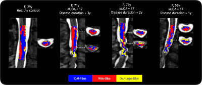
1612. Spinal
cord microstructure-based tissue classification in cervical
myelopathy
S. Balaji, S. Kolind, A. Traboulsee, A. MacKay, N. Dea
University of British Columbia, Vancouver, Canada
Impact: Cervical cord tissue was classified in people
with degenerative cervical myelopathy based only on
clustering quantitative MRI measures. Different proportions
of tissue clusters were seen in regions immediately above
compression sites compared to the entire imaged cord
(C2-C5).
|
|
 |
Computer Number: 84
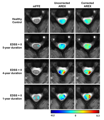
1613. Relaxation-compensated
CEST MRI in the spinal cord of multiple sclerosis patients at 3T
A. Cronin, G. Sweeney, L. Prock, D. Houston, I. Stuart, C.
McKnight, F. Bagnato, K. O'Grady, S. Smith
Vanderbilt University Medical Center, Nashville, United States
Impact: Initial results suggest that AREX shows
improvement over alternative methods and could offer
increased sensitivity to biochemical changes in the spinal
cord of MS patients.
|
|
 |
Computer Number: 85
1614. Ultra-high
field cervical spinal cord quantitative MRI: A 7T multi-center
study of traveling spines
V. Callot, M. Bennasser, S. Mchinda, D. Papp, R. Barry, L.
Beghini, A. Seifert, E. Alonso-Ortiz, J. Cohen-Adad, N.
Graedel, M. Callaghan, F. Eippert, N. Weiskopf, C. Aigner,
P. Freund, J. Vannesjo, A. Destruel, H. Dary, M. Guye, M.
Seif
CNRS/Aix-Marseille University, Marseille, France
Impact: Our multiparametric qMRI protocol for 7T spinal
cord imaging can enable multicenter clinical studies and
provide guidance for new investigators, ultimately advancing
diagnostic and prognostic capabilities for spinal cord
diseases while deepening our understanding of
neurodegenerative changes.
|
|
 |
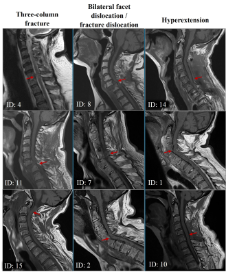
Computer Number: 86
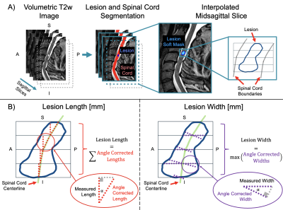
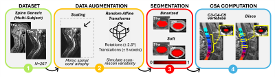
1615. Automatic
morphometry of spinal cord injury lesions
J. Valošek, D. Pfyffer, N. Karthik, L. Farner, S.
Schading-Sassenhausen, P. Freund, J. Cohen-Adad
Polytechnique Montreal, Montreal, Canada
Impact: Automatic computation of lesion morphometry can
replace manual measurements, thus facilitating large
multi-center studies in spinal cord injury patients by
reducing intra- and inter-expert variability and saving
time.
|
|
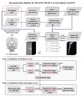 |
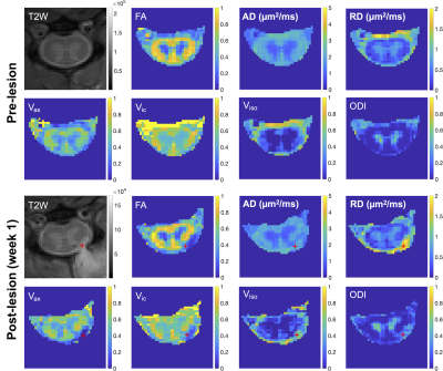
Computer Number: 87
1616. High-resolution
and High-fidelity DTI of Cervical Cord using 3D Reduced-FOV
Multiplexed Sensitivity Encoding (3D-rFOV-MUSE)
C. Yuan, S. Chen, L. Liang, X. Xu, H. Xiong, T. Liu, Y. Li,
N-K Chen, H-C Chang
The Chinese University of Hong Kong, Hong Kong, China
Impact: The proposed 3D-rFOV-MUSE technique can produce
high-fidelity csc-DTI at 1.0 mm-isotropic resolution, which
can precisely assess the microstructural integrity of the
cervical spinal cord. This may provide further
pathophysiological insights to aid differential diagnosis
for different cervical spinal cord diseases.
|
|
 |
Computer Number: 88
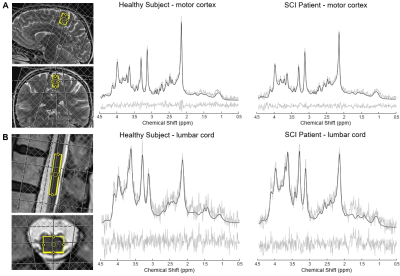
1617. Quantifying
Spinal Cord and Brain Metabolic Alterations in the Motor System
after Spinal Cord Injury Using Metabolite-Cycling Semi-Laser
¹H-MRS
A. Lebret, S. Schading-Sassenhausen, K. Şimşek, P. Gut, S.
Imhof, B. Zörner, R. Kreis, P. Freund, M. Seif
Balgrist University Hospital, Zurich, Switzerland
Impact: The feasibility of lumbar cord MRS has great
potential to assess tissue integrity non-invasively and
provide valuable insights into neurodegenerative processes,
with the potential for developing new biomarkers to improve
prognostication following SCI.
|
|
 |
Computer Number: 89
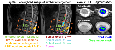
1618. Multi-echo
gradient echo MRI of the lumbosacral spinal cord reveals
level-dependent decreases in cross-sectional area in multiple
sclerosis
G. Dunay, F. Adepegba, A. Combes, A. Cronin, L. Narisetti,
G. Sweeney, L. Prock, D. Houston, A. Witt, X. Zhang, S.
Vandekar, F. Bagnato, S. Sriram, S. Smith, K. O'Grady
Vanderbilt University, Nashville, United States
Impact: Characterizing contributions of biological
variables to lumbosacral spinal cord MRI morphometry in
healthy controls enables detection of disease-related
effects such as cord and gray matter atrophy in pwMS,
informing future studies of imaging biomarkers in the
lumbosacral enlargement.
|
|
 |
Computer Number: 90
1619. Early-stage
structural and biochemical changes in cervical spinal cord after
sensory nerve root injury revealed by multi-parametric MRI
F. Wang, J. Gore, L. M. Chen
Vanderbilt University Medical Center, Nashville, United States
Impact: Multi-parametric MRI offers sensitive and
specific metrics for assessing changes within the spinal
cord after sensory nerve root injury. Our findings reveal
the early-stage structural and biochemical alterations at
the damaged nerve roots and the adjacent dorsal root entry
zone.
|
|
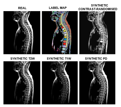 |
Computer Number: 91
1620. Generating
large-scale highly heterogenous synthetic MRIs for robust spinal
cord segmentation models
B. Brito Vega, P. Goebl, J. E. Iglesias, S. Narayanan, R.
Wolz, F. Barkhof, A. Eshaghi
University College London, London, United Kingdom
Impact: Our model paves the way for training
contrast-agnostic and resolution-independent MRI
segmentation models for spinal cord. This facilitates the
processing of routine care data supporting more robust,
translatable and generalisable models which can impact
patients with neurological disorders.
|
|
 |
Computer Number: 92
1621. Micro-
and Macrostructural Changes in the Brain and Spinal Cord in
Acute Spinal Cord Injury: A Multicenter qMRI Study
L. Farner, T. Emmenegger, M. Seif, A. Hug, N. Weidner, A.
Curt, P. Freund
Balgrist University Hospital, Zurich, Switzerland
Impact: By leveraging advanced Multiparameter Mapping
MRI techniques across 8 European centers, we have identified
specific volumetric and microstructural alterations that
correlate with functional outcomes. These findings
underscore the importance of early detection and targeted
interventions, potentially guiding future therapeutic
strategies.
|
|
 |
Computer Number: 93
1622. Comparison
of Acute In-vivo and Postmortem Ex-vivo MRI Metrics in Spinal
Cord Injury
N. Marini, N. Lesack, S. Morris, A. Yung, K. Bale, S.
George, A. Bauman, P. Kozlowski, Z. Samadi-Bahrami, C.
Fournier, P. Mattu, L. Parker, K. Dong, F. Streijger, W.
Moore, A. Velenosi, V. Hirsch-Reinshagen, B. Kwon, C. Laule
International Collaboration on Repair Discoveries, Vancouver, Canada
Impact:
Acute in-vivo MRI at time of spinal cord injury may be insufficient to predict the degree of permanent tissue damage. Additional MRI methods are needed to improve spinal cord injury long-term prognostication. |
|
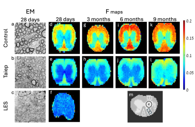 |
Computer Number: 94
1623. Magnetization
EXchange (MEX) MRI Reveals Myelin Content in ex-vivo Rat Spinal
Cord of Genetic Dysmyelination Mutants
E. Wilczynski, M. Teixeira Resende, B. August, I. Duncan, P.
Basser, Y. Cohen
Eunice Kennedy Shriver National Institute of Child health and Human Development (NICHD), National Institutes of Health (NIH), Bethesda, United States
Impact: The results validate the MEX sequence
capabilities to quantify myelin content, with the
prospective of clinical use. The main challenge is reducing
scan time. Additionally, the use of the Taiep model
shows great promise for further studying genetic
dysmyelination disorders.
|
|
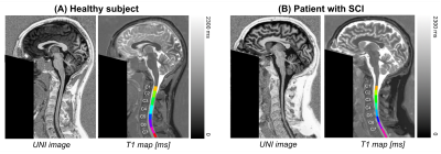 |
Computer Number: 95
1624. Simultaneous
T1 quantification across brain and entire cervical cord in
traumatic spinal cord injury (SCI)
A. Lebret, A. Forodighasemabadi, S. Schading-Sassenhausen,
P. Freund, V. Callot, M. Seif
Balgrist University Hospital, Zurich, Switzerland
Impact: T1 mapping
along the central nervous system enables better
understanding anterograde and retrograde degenerative
processes in vivo after SCI within scan times appropriate
for clinical routine.
|
|
 |
Computer Number: 96
1625. Minimum
sample size to detect spinal cord atrophy with automatic soft
segmentation
S. Bédard, E. Karthik, J. Valošek, J. Cohen-Adad
Polytechnique Montréal, Montréal, Canada
Impact: Reducing the required sample size will allow for
early spinal cord atrophy detection, especially in
multi-center and multi-contrast studies.
|
 Back to Meeting Home
 Back to the Program-at-a-Glance
The International Society for Magnetic Resonance in Medicine is accredited by the Accreditation Council for Continuing Medical Education to provide continuing medical education for physicians.
 View
Presentation Video
View
Presentation Video