Digital Poster
Segmentation Newborn/Fetal Brain
ISMRM & ISMRT Annual Meeting & Exhibition • 10-15 May 2025 • Honolulu, Hawai'i

Digital Poster
Segmentation Newborn/Fetal Brain
 |
Computer Number: 145
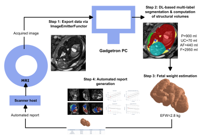
2405. Real-time
scanner-based automated fetal weight estimation and volumetry
reporting in fetal MRI
S. Neves Silva, A. Uus, S. McElroy, W. Norman, K. St Clair,
J. Aviles Verdera, S. Bansal, H. Waheed, J. Matthew, D.
Lloyd, J. Hajnal, L. Story, M. Rutherford, J. Hutter
King's College London, London, United Kingdom
Impact: Real-time fetal weight estimation and volumetry
using AI on the scanner enables faster and more individual
fetal MRI - hence paving the way for enhanced antenatal
diagnosis.
|
|
 |
Computer Number: 146
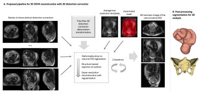
2406. Integrating
3D distortion correction in 3D deformable slice-to-volume
reconstruction of the whole uterus and pelvis for 0.55T T2w
fetal MRI
A. Uus, S. McElroy, A. Price, J. Aviles Verdera, S. Neves
Silva, S. Bansal, K. St Clair, M. Deprez, V. Kyriakopoulou,
L. Story, K. Colford, J. Hutter, M. Rutherford, J. Hajnal
King's College London, London, United Kingdom
Impact: This pipeline could be potentially useful for
various projects working on analysis of 3D large ROI
datasets.
|
|
 |
Computer Number: 147
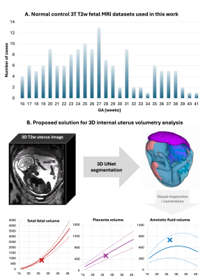
2407. Normative
volumetry growth models for the fetus, placenta and amniotic
fluid for 3D 3T T2w fetal MRI during 16 – 40 weeks GA range
A. Uus, M. Hall, A. Egloff Collado, C. Avena Zampieri, C.
Bradshaw, M. Deprez, J. Matthew, K. Colford, J. Hajnal, M.
Rutherford, J. Hutter, L. Story
King’s College London, London, United Kingdom
Impact: These models and segmentation network could be
potentially used in other research studies for normalisation
of fetal organ volumetry.
|
|
 |
Computer Number: 148
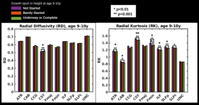
2408. The
hidden link between cerebral white matter development and
somatic height growth
J. Chad, C. Lebel
Baycrest Academy for Research and Education, Toronto, Canada
Impact: This work challenges the interpretation of WM
developmental trajectories as reflecting insular monotonic
growth. Instead, WM development appears to comprise not only
WM growth but also WM pruning that allocates energy for
somatic growth.
|
|
 |
Computer Number: 149
2409. Clinically
Integrated MRI-QSM Analysis & Reporting System to Establish
Brain Tissue Iron Levels in Pediatrics
H. Zamanian, E. Doyle, J. Wood, B. Tamrazi, M. Borzage, S.
Erberich
Children's Hospital Los Angeles, Los Angeles, United States
Impact: We developed an image processing orchestration
system that automates the identification, processing, and
reporting of QSM MRI data into PACS.
|
|
 |
Computer Number: 150
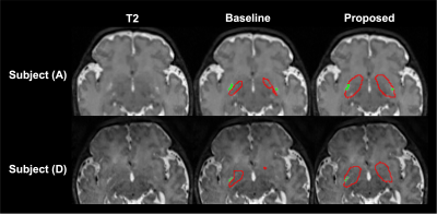
2410. Improving
Basal Ganglia Segmentation in Neonatal Brain for Dilated
Perivascular Space Assessment Using Soft Labels
D. Bak, J. Kang, Y. Nam, H. G. Kim
Hankuk University of Foreign Studies, Yongin, Korea, Republic of
Impact: The proposed soft label-based segmentation
method improves basal ganglia segmentation performance in
low-contrast neonatal MR images compared to
conventional hard label-based methods. The proposed method
could be beneficial for perivascular space assessment in
neonatal populations.
|
|
 |
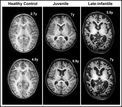
Computer Number: 151
2411. Differences
in Early Brain Structure Between Infants with Normal and
Abnormal Prognosis in External Hydrocephalus
h. zhao, y. sun, y. yin, x. li, m. wang, c. liu, j. yang, c.
jin
1.Department of Radiology, The First Affiliated Hospital of Xi'an Jiaotong University, Xi’an, China 2.Shaanxi Engineering Research Center of Computational Imaging and Medical Intelligence, Xi’an, China 3.Xi’an Key Laboratory of Medical Computational Imaging, Xi’an, China, Xi’an, China
Impact: Early MRI detection of cortical differences in
EH infants can guide interventions to prevent developmental
delays.
|
|
 |
Computer Number: 152
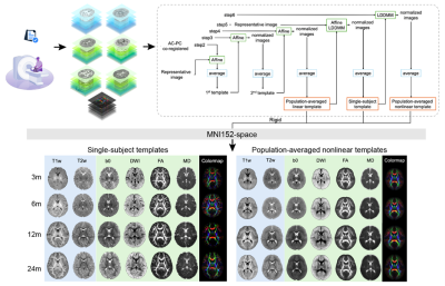
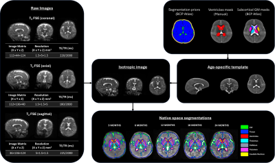
2412. Comprehensive
Multi-Modal MRI Templates of the Infant Brain: A Foundational
Resource for Early Developing Brain Studies
R. Li, R. Lin, S. Mophapatra, F. Wu, W. Wu, T. Zhu, C. E.
Li, S. Tan, K. Sindabizera, M. Ouyang, H. Huang
Children's Hospital of Philadelphia, Philadelphia, United States
Impact: The established foundational resource for infant
brain research offers dense and age-specific multi-modal MRI
templates. This resource empowers researchers to conduct
precise neuroimaging analyses of infants, unlocking insights
into brain structure, function, and connectivity in both
health and disease.
|
|
 |
Computer Number: 153
2413. MRI
brain volumetric analysis of type II GM1 gangliosidosis patients
treated with gene therapy
M. S. Shazeeb, C. Zoppo, J. Kolstad, J. Johnston, P.
D'Souza, A. Kuhn, Z. Vardar, A. Peker, A. Hader, R. King, H.
Celik, C. Lewis, C. Lindsay, Z. Rentiya, C. Lebel, S.
Vedantham, B. Vachha, M. Acosta, H. GrayEdwards, C. Tifft
University of Massachusetts Chan Medical School, Worcester, United States
Impact: Our study addressed the need for quantitative
neural biomarkers in type II GM1 gangliosidosis which
correlated with clinical markers. Through longitudinal brain
volumetric analysis using MRI, we demonstrated the efficacy
of gene therapy in monitoring disease progression/regression
in GM1 patients.
|
|
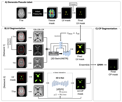 |
Computer Number: 154
2414. Automatic
Lateral Ventricle and Choroid Plexus Segmentation method in
infant brain MR images
J. Kang, H. G. Kim, Y. Nam
Hankuk university of Foreign Studies, Yongin-si, Korea, Republic of
Impact: Our CP and LV segmentation method provides
improved performance in infant MRI , demonstrating the
potential for more robust quantitative analysis in the
infant population. This could help to explore the
relationship between glymphatic functions and the early
stage of neurodevelopment.
|
|
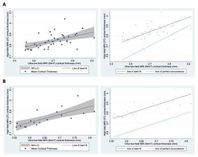 |
Computer Number: 155
2415. Investigating
cortical thickness in children on ultra-low-field and high-field
MRI
C. Wedderburn, N. Bourke, L. Bradford, J. Ringshaw, T.
Malaba, H. Theunissen, S. Williams, L. Davel, J. Read, H.
Reynolds, N. He, A. Colbers, D. Wang, S. Khoo, L. Myer, K.
Donald
University of Cape Town, Cape Town, South Africa
Impact: Paediatric cortical thickness measurements may
be obtained using ultra-low-field MRI that correspond with
high-field MRI and improve with scan quality. Further work
is needed to advance ultra-low-field acquisition and
processing pipelines to optimise cortical thickness and
assess other cortical metrics.
|
|
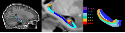 |
Computer Number: 156
2416. Hippocampal
morphometry is altered in children with congenital heart disease
B. Gal-Er, A. Bonthrone, M. van der Meijden, A. Chew, C.
Casella, Y. Brackenier, M. Cleri, P. Di Cio, A. Elgoff, K.
Pushparajah, J. Simpson, M. Rutherford, A. D. Edwards, S.
Malik, L. Cordero-Grande, J. Hajnal, C. Nosarti, J.
O’Muircheartaigh, S. Counsell
King's College London, London, United Kingdom
Impact:
This study extends previous work showing reduced hippocampal volume in this population by identifying reduced hippocampal gyrification and subfield volume in children with CHD. Altered hippocampal development may be a key determinant of neurodevelopmental impairments observed in children with CHD. |
|
 |
Computer Number: 157
2417. Diagnostic
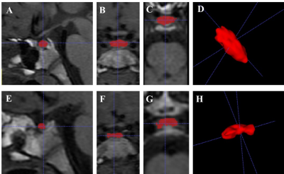
value of accurately MRI adenohypophyseal volume in evaluation of
HPG axis activation in pre- and at-puberty children
L. Qin, W. Liu, L. Dong
Tongji Hospital, Tongji Medical College, Huazhong University of Science and Technology, Wuhan, China
Impact: aPV and aPH demonstrated potential diagnostic
value in assessing HPG-axis initiation status. The multiple
linear regression equations incorporating age, weight and
adenohypophysis volume showed promise for predicting LH peak
and LH/FSH ratio, providing insights into non-invasive
methods for assessing precocity.
|
|
 |
Computer Number: 158
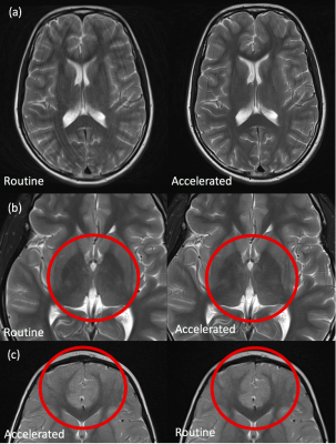
2418. Impact
of AI Acceleration on Image Quality and Diagnostic Quality in
Clinical Paediatric Neuroimaging
T. McGeown, J. Cleary, S. Kafiabadi, N. Adroja, N. Mellor,
T. Moon, C. O'Brien, S. Shah
King's College Hospital NHS Foundation Trust, London, United Kingdom
Impact: Accelerated imaging techniques can be used to
significantly reduce scan times in paediatric brain imaging
whilst maintaining image quality, supporting confident
clinical deployment.
|
|
 |
Computer Number: 159
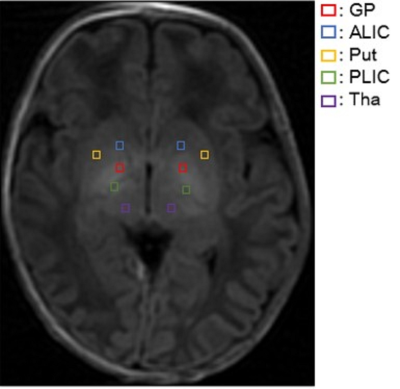
2419. Association
of T1WI Abnormality with the Interval between MRI and
Measurement of Bilirubin Level Peak on Hyperbilirubinemia
Neonates
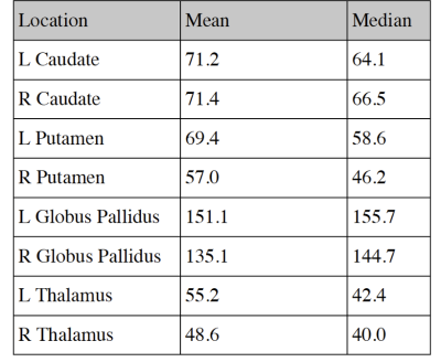
R. Wang, H. Tian, Z. Ren, K. Zhang, X. Li, X. Zheng, X. Li
Department of Radiology, the First Affiliated Hospital of Xi’an Jiaotong University, Xi’an, China
Impact: Abnormal T1WI signal changes in basal ganglia
nuclei of hyperbilirubinemic neonates vary with MRI timing.
This insight aids clinicians to avoid performing MRI scans
on subjects when the time is suboptimal, which may lead to
false negative results on T1WI.
|
|
 |
Computer Number: 160
2420. MiniMORPH:
A Morphometry Pipeline for Low-Field MRI in Infants
C. Casella, N. Bourke, A. Leknes, A. Zahra, D. Scheiene, R.
Macleod, J. Cole, F. Biondo, M. Zabihi, V. Nankabirwa, K.
Donald, M. Bruchhage, J. O'Muircheartaigh
King's College London, London, United Kingdom
Impact: We provide a scalable, open-source solution for
segmentation of ULF MRI infant data, unlocking new
possibilities for assessing neurodevelopment in diverse
settings.
|
 Back to Meeting Home
 Back to the Program-at-a-Glance
The International Society for Magnetic Resonance in Medicine is accredited by the Accreditation Council for Continuing Medical Education to provide continuing medical education for physicians.
 View
Presentation Video
View
Presentation Video