Digital Poster
Mid-Field Applications
ISMRM & ISMRT Annual Meeting & Exhibition • 10-15 May 2025 • Honolulu, Hawai'i

 |
Computer Number: 113
1642. A
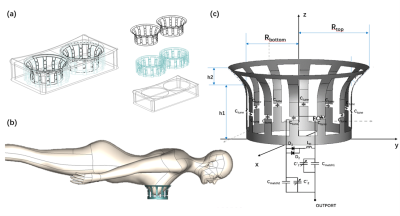
flowerpot-shaped birdcage coil designed for breast MRI at 0.35T
X. Song, Q. Liu, Z. Jiang, C. Lin, B. Qiu
Institute of Advanced Technology, University of Science and Technology of China, Hefei, Anhui, China
Impact: We proposed a novel flowerpot-shaped birdcage
coil for breast MRI using metamaterials in a 0.35T system,
resulting in an improved signal-to-noise ratio (SNR) and
receiving sensitivity(RS).
|
|
 |
Computer Number: 114
1643. Resting
state functional MRI with Multi echo GRE EPI on a high
performance 0.5 T Head-only Scanner
A. Halder, S. Chavez, C. Harris, C. Wiens, A. Curtis
Western University, Mississauga, Canada
Impact: ME-EPI is feasible for producing reliable
rs-fMRI at 0.5 T, potentially expanding access of rs-fMRI in
the acute setting.
|
|
 |
Computer Number: 115
1644. Lumbar
spine MRI at 0.6T
M. Nagtegaal, Y. Dong, E. Ercan, P. Börnert, A. Webb, A.
Webb, M. van Osch, K. van Langevelde
Leiden University Medical Center, Leiden, Netherlands
Impact: This study shows that a complete lumbar MRI
protocol can be acquired at 0.6T around 11 minutes of
acquisition time, paving the way for future studies in
patients, with a special focus for imaging around implants.
|
|
 |
Computer Number: 116
1645. Advanced
AI Optimized 16-Channel Modular Coil for Enhanced Flexibility in
Low-Field MRI
S. Kumar, R. Stormont, J. Wild, F. Robb, R. Venkatesan
University of Sheffield, Sheffield, United Kingdom
Impact: A lightweight, flexible 16-channel modular coil
scanned multiple anatomies—including spine, knee, abdomen,
pelvis, shoulder, lung, neck, foot, and ankle on a 0.5T
system. Its elements were strategically placed for optimal
parallel imaging acceleration across various orientations.
|
|
 |
Computer Number: 117
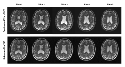
1646. Towards
synthetic T2-weighted imaging based on multiple acquisition
bSSFP at 0.55T
K. Keskin, B. Li, K. Nayak
University of Southern California, Los Angeles, United States
Impact: We demonstrate synthetic T2-weighted imaging
based on multiple bSSFP acquisitions, and voxel-wise
estimation of NMR parameters. This could be useful for
relaxometry and synthetic imaging at mid and low-field
strengths where bSSFP performance is favorable.
|
|
 |
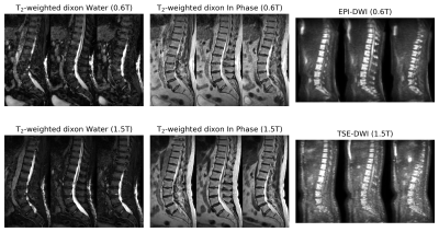
Computer Number: 118
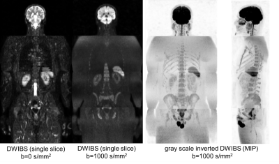
1647. Whole
body DWIBS and Water-Fat resolved Dixon for Midfield MRI
Y. Dong, M. Nagtegaal, E. Ercan, J. Smink, K. van
Langevelde, B. Boekestijn, A. Webb, M. J. van Osch, P.
Börnert
C.J. Gorter MRI Center, Department of Radiology, LUMC, Leiden, Netherlands
Impact: This study demonstrates that midfield MRI can
achieve clinically viable whole-body DWIBS and Dixon
imaging. These findings could encourage broader clinical use
of midfield MRI, particularly for patients with implants or
susceptibility to high-field MRI risks.
|
|
 |
Computer Number: 119
1648. Field
Strength Dependency of Geometric Distortion in
Diffusion-Weighted single-shot EPI: Comparing 0.6 T and 1.5 T
Scanners
Y. Dong, M. Nagtegaal, E. Ercan, J. Smink, H. Peeters, K.
van Langevelde, B. Boekestijn, A. Webb, P. Börnert, M. J.
van Osch
C.J. Gorter MRI Center, Department of Radiology, LUMC, Leiden, Netherlands
Impact: 0.6T ss-EPI DWI offers distortion-free imaging
in anatomies prone to artifacts at higher fields, such as
the prostate, brain, and spine. This approach could enhance
diagnostic accuracy, improve patient comfort, and support
broader clinical adoption of midfield MRI.
|
|
 |
Computer Number: 120
1649. The
TwinsUK MR Imaging study protocol: Brain and spine at 3T and
cardiac plus whole-body at 0.55T
R. Thornley, Z. Ning, L. S. Canas, J. Cleary, P. Bridgen, P.
Di Cio, M. Cleri, A. Kaushal, S. Jeljeli, P. G. Masci, M.
Niglas, B. Whitcher, M. Modat, J. Bell, E. Thomas, J.
Maynard, V. Goh, A. Isaac, S. Giles, C. Steves, S. Ourselin,
A. Chiribiri, J. Hajnal, A. Price
King's College London, London, United Kingdom
Impact: This study will generate a comprehensive MRI
resource in a twin cohort of ~2500 participants. Combined
with biological data, this facilitates the study of ageing
and has potential to lead to more personalised approaches to
managing health as we age.
|
|
 |
Computer Number: 121
1650. Experience
from planning and operationalising a 0.55 T MRI system in a
resource-constrained setting
D. Kandasamy, L. Lokesh, S. Gamanagatti, A. Goyal, Y.
Sharma, B. Schmitt, P. Misra, R. Kumar, H. Salve, R. Ramb,
R. Kaur, V. Gulani, R. Sharma
All India institute of Medical Sciences, New Delhi, New Delhi, India
Impact: This study demonstrates that low-field MRI can
meet essential diagnostic needs in rural settings, reducing
travel and enabling timely care. Findings support the
broader adoption of 0.55T MRI to improve access and early
detection of actionable findings in resource-limited areas.
|
|
 |
Computer Number: 122
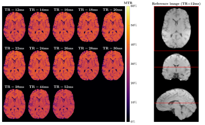
1651. Optimized
contrast-to-noise ratio efficiency of Magnetization Transfer
brain imaging at 0.55T
D. Leitão, D. West, S. McElroy, R. Tomi-Tricot, J. Hajnal,
T. Wood, S. Malik
King's College London, London, United Kingdom
Impact: Efficient acquisitions at lower field resolves
the two greatest difficulties of deploying Magnetization
Transfer Ratio images in the clinic, namely scan time and
SAR concerns.
|
|
 |
Computer Number: 123
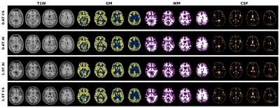
1652. Characterizing
differences between white and gray matter T1W-based
segmentations at 0.6T and 1.5T
N. Jabarimani, E. Ercan, Y. Dong, N. Pezzotti, A. Webb, P.
Börnert, M. Staring, M. van Osch, M. Nagtegaal
Leiden University Medical Center, Leiden, Netherlands
Impact: This study confirms that mid-field MRI (0.6T) T1-weighted
images can be used for white and gray matter segmentation
with reproducible volume measures. These findings may enable
longitudinal monitoring of brain volume and inspire future
research into improved segmentations.
|
|
 |
Computer Number: 124
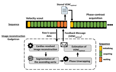
1653. Automatic
velocity encoding (VENC) calibration for accurate quantitative
flow measurement
P. Daude, R. Ramasawmy, A. Javed, D. Franson, K. Chow, A.
Campbell-Washburn
National Heart, Lung & Blood Institute, Bethesda, United States
Impact: Inline automatic velocity encoding calibration
ensures the optimal precision in flow measurements and
simplifies the acquisition workflow.
|
|
 |
Computer Number: 125
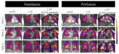
1654. Comparison
of Lung Ventilation and Perfusion on Commercial 0.55T and 1.5T
Systems
J. Varghese, K. Binzel, Y. Liu, O. Bieri, O. Simonetti, G.
Bauman
The Ohio State University, Columbus, United States
Impact: The comparative performance of functional lung
MRI using matrix pencil decomposition for estimation of
regional fractional ventilation and perfusion on commercial
0.55T and 1.5T systems is demonstrated, highlighting the
advantages of low field MRI for pulmonary imaging.
|
|
 |
Computer Number: 126
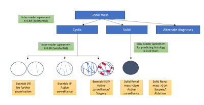
1655. Clinical
Utility of a Commercial 0.55 Tesla MRI System for the Evaluation
of Renal Lesions
R. Rajeev, T. Lin, H. Hussain, S. Wells, W. Weadock, A.
Ramachandran, M. Masotti, B. Mervak, A. Aslam, R. Chahine,
B. Hirshberg, J. Richardson, M. Masotti, N. Seiberlich, V.
Gulani, M. Mendiratta-Lala
University of Michigan , Ann Arbor, United States
Impact: 0.55T MRI can effectively characterize solid and
cystic renal masses and risk-stratify cystic renal masses
according to the Bosniak classification without compromising
diagnostic accuracy.
|
|
 |
Computer Number: 127
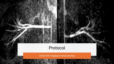
1656. Renal
Artery Embolization Under 0.55T: Real-Time Navigation,
Visualization, Segment Selection, and Perfusion
N. Ooms, E. Brandner, J. Roll, R. Anderson, J. Krieger, P.
Sutphin, S. Kalva
Purdue University, West Lafayette, United States
Impact: The speed of procedure, as well as visual
confirmation in real time, has led us to continue
investigating Renal Artery Embolization techniques under MRI
guidance. We will also be investigating other procedures
that would benefit from real time MRI guidance.
|
|
 |
Computer Number: 128
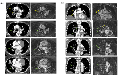
1657. Detectability
of Pulmonary Embolism by contrast-free MR Angiography at 0.55T
R. Rizzo, R. Rajeev, J. Richardson, Y. Sharma, M. Stanzione
Galizia, C. Fung, V. Gulani, N. Seiberlich
University of Michigan, Ann Arbor, United States
Impact: Whole-body 0.55T MR scanners, with reduced
air-tissue interface susceptibility, offer a preliminary
promising alternative to CTPA for detecting pulmonary
embolism (PE) in patients for whom CTPA is unsuitable,
potentially enabling non-contrast PE detection in
high-volume clinical settings.
|
 Back to Meeting Home
 Back to the Program-at-a-Glance
The International Society for Magnetic Resonance in Medicine is accredited by the Accreditation Council for Continuing Medical Education to provide continuing medical education for physicians.
 View
Presentation Video
View
Presentation Video