Digital Poster
AI-Powered Analysis in Neuroimaging
ISMRM & ISMRT Annual Meeting & Exhibition • 10-15 May 2025 • Honolulu, Hawai'i

Digital Poster
AI-Powered Analysis in Neuroimaging
 |
Computer Number: 17
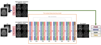
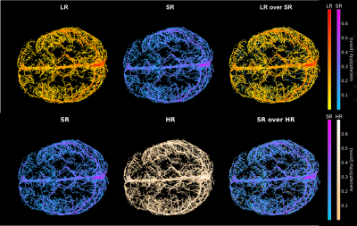
4285. Improving
Low-Angular Resolution Diffusion MRI with 3D Deep Learning: A
Model Assessment
N. Tucksinapinunchai, S. Angkurawaranon, R. Boonsuth, U.
Yarach
Chiang Mai University, Chiang Mai, Thailand
Impact: The improved quality and reliability of the
diffusion parametric maps produced by our trained 3D-DL
model may be advantageous for clinical applications or the
investigation of white matter microstructure in various
demographics, including transgender individuals, in future
research.
|
|
 |
Computer Number: 18
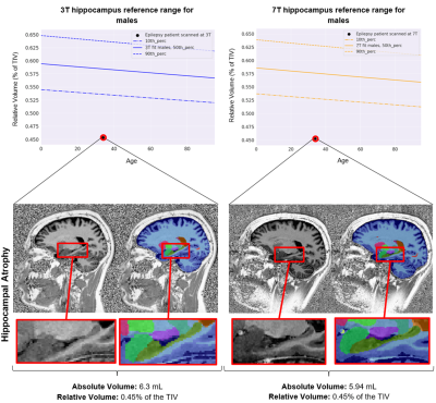
4286. Advancing
Brain Morphometry at 7T: A Pilot Study on Epilepsy Patients
L. Bacha, T. Di Noto, P. B. Venkategowda, K. Prabhu, G. F.
Piredda, G. Bonanno, J. Slotboom, D. Seiffge, M. Goeldlin,
R. Hoepner, S. Vulliemoz, M. Seeck, K. Schindler, M. Baud,
J-P Thiran, T. Kober, T. Hilbert, P. A. Liebig, R.
Heidemann, R. Wiest, P. Radojewski, B. Maréchal
Siemens Healthineers International AG, Lausanne, Switzerland
Impact: This work introduces a novel and reliable brain
morphometry algorithm that provides detailed structural
insight for enhanced clinical decision support in epilepsy
care.
|
|
 |
Computer Number: 19
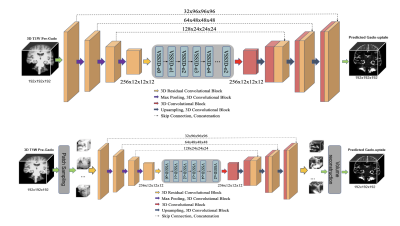
4287. Deep
learning of MRI contrast enhancement for mapping cerebral blood
volume from single-modal non-contrast scan with Mamba3D-CNN
hybrid model
Y. Zhang, A. Cao, V. Rao, J. Guo
Columbia University, New York, United States
Impact: By accurately estimating cerebral blood volume,
this approach eliminates risks associated with gadolinium
administration, such as long-term tissue retention. This
advancement enables functional imaging for researchers and
clinicians, providing a safe and cost-effective alternative
for studying and diagnosing neurodegenerative diseases.
|
|
 |
Computer Number: 20
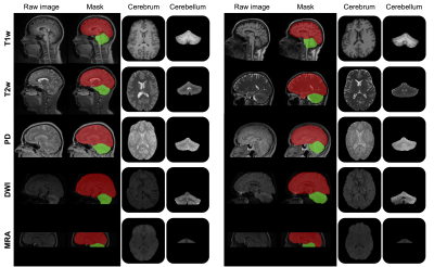
4288. Joint
Extraction of Cerebrum and Cerebellum from Lifespan MRIs
L. Wang, Y. Sun, G. Li, W. Lin, L. Wang
UNC-Chapel Hill, Chapel Hill, United States
Impact: Our method unifies cerebrum and cerebellum
extraction, addressing anatomical differences, reducing time
complexity, and ensuring accuracy across modalities and
ages. This enhances neurological research and clinical
diagnostics by enabling precise analysis and monitoring of
brain structures.
|
|
 |
Computer Number: 21
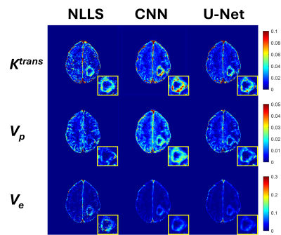
4289. Three-Point
Deep Learning Framework for Protocol-Independent and AIF-Free
DCE-MRI Parameter Estimation in Gliomas
P. Prajapati, A. Kandpal, S. Srivastava, R. Gupta, A. Singh
Indian Institute of Technology Delhi, New Delhi, India
Impact: The proposed DL approach enables robust DCE-MRI
quantification in gliomas using minimal temporal sampling,
eliminating AIF dependencies while maintaining accuracy in
substantially less time. Facilitating multi-center clinical
adoption and efficient pre-operative tumor characterization
and treatment monitoring.
|
|
 |
Computer Number: 22
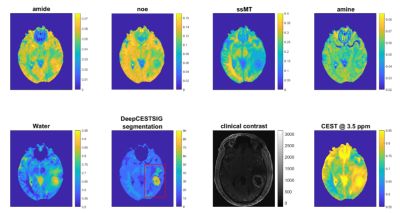
4290. DeepCESTSig:
Brain and Tumor Subregion Delineation via Chemical Exchange
Saturation Transfer (CEST) MRI
J. R. Rajput, M. S. Fabian, T. A. moehle, A. Mennecke, M.
Schmidt, A. Dörfler, A. Maier, M. Zaiss
Universitätsklinikum Erlangen, Erlangen, Germany
Impact: Segmentation based on CEST signatures improves
the delineation of brain tissue and identification of
tumors, enabling better clinical decision-making. The
approach improves neuroimaging techniques by using
biochemical contrasts and can improve the results in the
diagnosis of malignant brain areas.
|
|
 |
Computer Number: 23
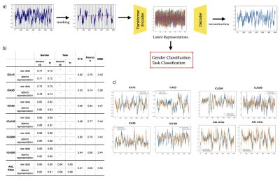
4291. Task-Agnostic
Brain Representations: A Foundation Model for fMRI Using Masked
Autoencoders
M. Ferrante, S. Iervese, L. Astolfi, N. Toschi
University of Rome Tor Vergata, Rome, Italy
Impact: We foundation model for fMRI, trained on
resting-state data from the HCP to develop generalizable
brain representations. Using self-supervised learning, this
task-agnostic model can be applied to various neuroscience
tasks, including physiological prediction and brain
decoding.
|
|
 |
Computer Number: 24
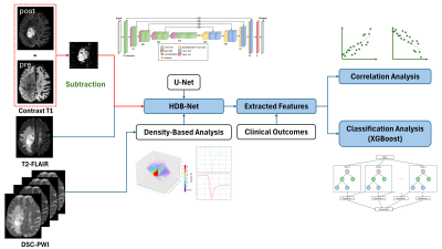
4292. Investigating
Prognostic Value of Dynamic Susceptibility Contrast Perfusion
MRI-Derived Features for Glioblastoma Survival by Deep Learning
L. Tang, Q. Gu, T. Wu, A. Goldman-Yassen, H. Mao
Emory University, Atlanta, United States
Impact: Using our Hierarchical Density-Based Network
(HDBNet) to investigate hemodynamic information in dynamic
susceptibility contrast perfusion-weighted imaging (DSC-PWI)
reveals key features that can enhance GBM prognosis,
supporting the importance of including hemodynamic and
physiological imaging data in future GBM research
|
|
 |
Computer Number: 25
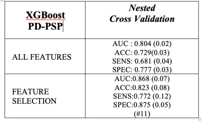
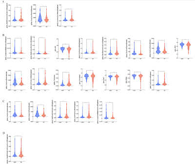
4293. Investigate
brain volumes with machine learning algorithms to differentiate
PD and PSP patients
C. Calomino, M. Bianco, A. Quattrone
Magna Graecia Univerisity, Catanzaro, Italy
Impact: This study provides first evidence of
alterations in subcortical volume and cortical thickness
between Parkinson’s disease patients and Progressive
Suplanuclear Palsy patients using a rigorous approach
combining nested cross validation, XGBoost and SHAP as
feature selection.
|
|
 |
Computer Number: 26
4294. Radiomics
features reveal the peritumoral heterogeneity of glioblastoma
L. Rui, A. Kai, Z. Jing
Lanzhou University Second Hospital, Lanzhou, China
Impact: The imaging features can provide objective
evidence for the peritumoral heterogeneity of GBM and MT,
and provide help for the clinical treatment of patients.
|
|
 |
Computer Number: 27
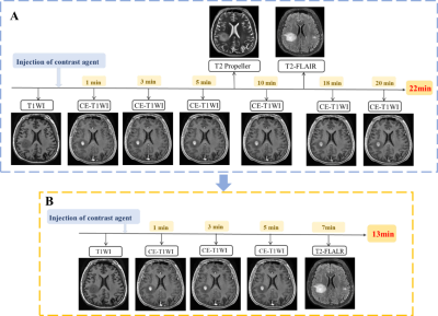
4295. The
effect of time-delayed contrast-enhanced T2-FLAIR on the
visualization of large-volume brain metastases
S. Du, Y. Yin, D. Pylypenko, G. Gong
Shandong Cancer Hospital, Jinan, China
Impact: Our results show that combining time-delayed CE
T2-FLAIR and CE T1WI enhances BM visualization and GTV
segmentation accuracy, allowing quantitative analysis of BM
imaging differences in various sequences through GTV volume
and shape evaluation.
|
|
 |
Computer Number: 28
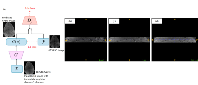
4296. BOLD
Acquisitions and GAN Synthetized VASO Contrasts for Rapid
Layer-dependent fMRI
A. Saxena, D. Bharti, T. B. D. Yeo, A. Ajala
GE Healthcare, Bangalore, India
Impact: We present a method to eliminate the need for
implementing VASO pulse sequence by synthetically generating
VASO images from acquired BOLD images.
|
|
 |
Computer Number: 29
4297. Leveraging
transfer learning for the super-resolution reconstruction of QSM
with limited data for the study of the cerebrovasculature
S. Zappalà, E. Patitucci, I. Driver, D. Gallichan, R. Wise,
M. Germuska
Cardiff University, Cardiff, United Kingdom
Impact: By demonstrating the effectiveness of transfer
learning with a 3D Densely Connected Super-Resolution
Network (DCSRN) model, this study provides a practical
approach for researchers to improve the resolution of their
own QSM data, even with limited resources.
|
|
 |
Computer Number: 30
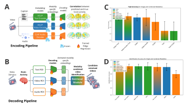
4298. Cross-modal
brain decoding: using fMRI to decode video stimuli from
integrated sensory streams
M. Ferrante, T. Boccato, N. Toschi
University of Rome Tor Vergata, Rome, Italy
Impact: This work opens pathways for more accurate brain
decoding in multisensory contexts, potentially advancing
brain-computer interfaces and aiding clinical applications
in sensory processing disorders.
|
|
 |
Computer Number: 31
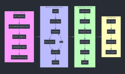
4299. Enhancing
3D Brain MRI Using Super-Resolution through U-Net Architecture
S. Singh, B. V. R. Kumar, S. Pathak, R. Jha, M. Singh, A.
Parihar, B. Ojha, C. Srivastava, D. Dwivedi
King George's Medical University, Lucknow, India
Impact: Utilizing SR for 3D MRI images is uncommon, yet
it significantly enhances MRI efficiency and resolution. The
proposed architecture reduces computational costs while
improving results, facilitating quicker MRI execution
without compromising image quality.
|
|
 |
Computer Number: 32
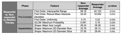
4300. Predictors
of Ablation Therapy Response in Moderately Differentiated HCC
using Radiomics Features from Multi-phasic DCE-MRI
X. Li, Z. Mohammadigolda, Q. Miao, P. Keshavarz, A. Suri, J.
Chiang, K. Sung, D. Lu
University of California, Los Angeles, Los Angeles, United States
Impact: This study identifies radiomics shape and
texture-based features from multiphase MRI that
differentiate responders from non-responders to thermal
ablation in moderately differentiated HCC. These features
offer insights into quantitative biomarkers, emphasizing
the role of imaging in optimizing therapy.
|
 Back to Meeting Home
 Back to the Program-at-a-Glance
The International Society for Magnetic Resonance in Medicine is accredited by the Accreditation Council for Continuing Medical Education to provide continuing medical education for physicians.
 View
Presentation Video
View
Presentation Video