Digital Poster
Deep Learning for Image Enhancement: Part II
ISMRM & ISMRT Annual Meeting & Exhibition • 10-15 May 2025 • Honolulu, Hawai'i

Digital Poster
Deep Learning for Image Enhancement: Part II
 |
Computer Number: 17
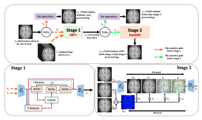
3830. QG-MoCo:
Quality-Guided Coarse- and Fine-Grained Path Selection For MRI
Motion Correction
F. Li, Z. Zhou, Y. Fang, J. Cai, Q. Wang
ShanghaiTech Univerisity, Shanghai, China
Impact: The quality-guided MoCo method effectively
reduces 3D motion artifacts following the automatic
selective path in different granularity for progressive
correction. Emphasizes the benefits on model accuracy and
efficiency by taking MoCo operations based on the routing
strategy with quality consideration.
|
|
 |
Computer Number: 18
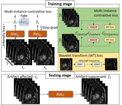
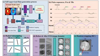
3831. Self-supervised
multi-instance contrastive learning for reduction of cardiac
bSSFP off-resonance artifacts
Z. Chen, Y. Emu, J. Gao, H. Chen, X. Tang, C. Hu
Shanghai Jiao Tong University, Shanghai, China
Impact: Obtaining corrupted-clean bSSFP image pairs is
challenging, particularly with cardiac devices or high-field
MR. Our proposed self-supervised approach mitigates
off-resonance artifacts and provides a practical solution
for reliable bSSFP cine imaging.
|
|
 |
Computer Number: 19
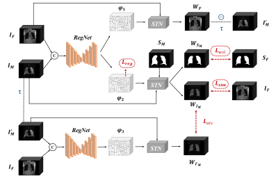
3832. Appearance
Transformation Consistency Network for Registering 1H MRI and
129Xe MRI of the lungs
Z. Bao, S. Xiao, Z. Chen, X. Zhou
State Key Laboratory of Magnetic Resonance and Atomic and Molecular Physics, National Center for Magnetic Resonance in Wuhan, Wuhan Institute of Physics and Mathematics, Innovation Academy for Precision Measurement Science and Technology, Chinese Academy of Sciences–Wuhan National Laboratory for Optoelectronics, Huazhong University of Science and Technology, 430071, wuhan, China
Impact: Our results will impact radiologists and
researchers by enabling precise lung image registration,
facilitating new investigations into lung pathologies.
|
|
 |
Computer Number: 20
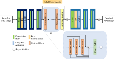
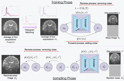
3833. Unsupervised
Denoising Method for Multi-sequence Low-field MRI in Veterinary
Imaging
J. Tang, W. Zu, J. Liu, C. Jin, Z. Zhang
School of Biomedical Engineering, Shanghai Jiao Tong University, Shanghai, China
Impact: In the absence of reference images, our method
preserves the structural features of images while
effectively reducing noise. It achieve noise reduction for
low-field images in a shorter time and has great potential
for improving low-field image quality improvement.
|
|
 |
Computer Number: 21
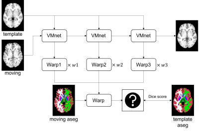
3834. FuseMorph:
accurate and time-efficient MRI 3D T1 Image deformable
registration with iterative search and deep learning
P-M Sun, T-Y Huang, T-C Chuang, Y-R Lin, H-W Chung
Department of Electrical Engineering, National Taiwan University of Science and Technology, Taipei, Taiwan
Impact: This
method improves MRI alignment accuracy and accelerates
processing, offering a reliable tool for both research and
clinical applications. It also enhances downstream tasks,
such as VBM analysis, allowing them to be performed with
greater speed and precision.
|
|
 |
Computer Number: 22
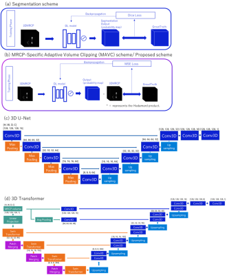
3835. MRCP-Specific
Adaptive Volume Clipping: A Deep Learning Method for Automated
Removal of Unnecessary Areas in MRCP images
Y. Sugimoto, N. Fujita, S. Funayama, S. Ichikawa, S.
Goshima, Y. Terada
University of Tsukuba, Tsukuba, Japan
Impact: The MRCP-Specific Adaptive Volume Clipping
method enhances MRCP examination efficiency by automatically
removing unnecessary regions in maximum-intensity projection
images of MRCP. This approach reduces manual workload,
offering flexibility without precise segmentation, and
improves workflow and diagnostic accuracy.
|
|
 |
Computer Number: 23
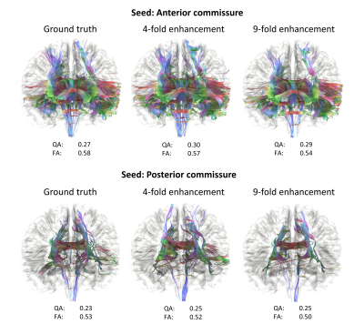
3836. Optimizing
feature-based loss functions for AI Super-resolution of 7T Brain
Diffusion MRI
D. Lohr, R. Werner
University Hospital Hamburg-Eppendorf, Hamburg, Germany
Impact: Our results show how feature-based loss
functions need to be adapted to work well for SR models
targeting MR diffusion data. We further demonstrate how such
SR models may be trained using publicly available data,
enabling reproducibility and application.
|
|
 |
Computer Number: 24
3837. Accelerated
EPR Imaging Using Deep Learning Denoising
I. Canavesi, N. Viswakarma, B. Epel, A. McMillan, M. Kotecha
O2M Technologies, LLC, Chicago, United States
Impact: EPR images with physically enhanced deep
learning techniques improve image SNR and reduce artifacts.
This advancement can be translated to reduce acquisition
time, reduce deposited power, and enable large object oxygen
imaging, bringing EPRI one step closer to clinical
translation.
|
|
 |
Computer Number: 25
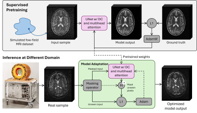
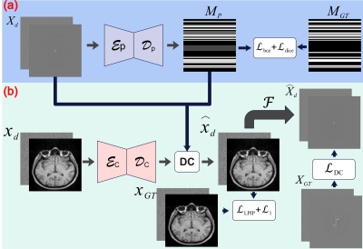
3838. Deep
Learning Image Denoising for In-Vivo Low-Field MRI Using
Test-Time Training
D. Schote, C. Kolbitsch, L. Winter, A. Kofler
Physikalisch-Technische Bundesastalt (PTB) Braunschweig und Berlin, Berlin, Germany
Impact: Noise-free reference data for supervised
training of low-field MRI denoising models does not exist.
Using supervised pretraining on simulated data combined with
self-supervised test-time training narrows the performance
gap in low-field MRI denoising models when training and
testing data differs.
|
|
 |
Computer Number: 26
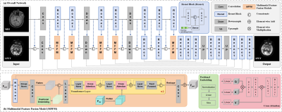
3839. Multimodal
Feature-Guided Diffusion Model for Low-Dose PET Image Denoising
with MRI
Y. Jin, G. Lin, H. Liu, C. Zhou, X. Zhang, W. Fan, N. Zhang,
H. Zheng, D. Liang, P. Cao, Z. Hu
Shenzhen Institute of Advanced Technology, Chinese Academy of Sciences, Shenzhen, China
Impact: The research can enhance the quality of low-dose
PET imaging and reduce the radiation risk for patients, and
it also inspires more feasible imaging solutions for the
global health field, holding significant scientific and
clinical importance.
|
|
 |
Computer Number: 27
3840. Denoising
7T Structural MRI with Conditional Generative Diffusion Models
B. Li, Y. Wang, Y. Liang, M. Carlson, P. DiGiacomo, H. Moein
Taghavi, J. Maclaren, M. Bell, E. Mormino, V. Henderson, G.
Zaharchuk, B. Rutt, W. Shao, M. Georgiadis, M. Zeineh
Stanford University, Palo Alto, United States
Impact: Our newly introduced 7T Conditional Diffusion
Model (7TCDM) enables faster MRI acquisition by providing
high-quality denoised images from shorter scans, increasing
the feasibility of scanning patients in shorter times while
preserving essential anatomical details.
|
|
 |
Computer Number: 28
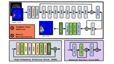
3841. AI-Enhanced
Super-Resolution for Metabolite MRI Imaging
E. Bjørkeli, J. T. Geitung, M. Esmaeili
Akershus University Hospital, Lørenskog, Norway
Impact: This improved SR approach significantly enhances
metabolite map quality, offering clinicians a valuable tool
for detailed neurological assessment.
|
|
 |
Computer Number: 29
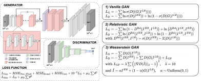
3842. Applications
of generative adversarial networks for super-resolution of
cerebrovascular 4D Flow MRI
O. Welin Odeback, E. Ferdian, A. Young, J. Schollenberger,
C. A. Figueroa, T. Granberg, A. Fyrdahl, D. Marlevi
Karolinska Institute, Stockholm, Sweden
Impact: This study highlights the potential of
generative adversarial networks to enhance super-resolution
in 4D Flow MRI, enabling more accurate intracranial flow
assessments near vessel walls.
|
|
 |
Computer Number: 30
3843. Partial
volume estimation from MRF acquisition using a deep learning
approach
T. Ding, Y. Gao, Z. Xiong, F. Liu, M. Cloos, H. Sun
The University of Queensland, Brisbane, Australia
Impact: This study's self-supervised deep learning
approach for partial volume estimation directly from MRF
signals could improve diagnostic accuracy and streamline
quantitative MRI processes. It opens avenues for real-time,
artifact-resilient tissue characterization, potentially
transforming clinical workflows and supporting
patient-specific imaging studies.
|
|
 |
Computer Number: 31
3844. A
Physics-Informed Deep Learning Method for Correcting Motion
Artifacts in Brain MR Imaging
M. Safari, Z. Eidex, M. Hu, C-w Chang, R. L. Qiu, T. Liu, T.
Liu, H. Mao, X. Yang
Emory University, Atlanta, United States
Impact: Our physics-informed deep learning model
markedly reduces motion artifacts in MRI scans, enhancing
image quality. By minimizing the need for repeat scans, this
method could significantly decrease healthcare costs and
bolster the reliability of downstream MR imaging
applications.
|
|
 |
Computer Number: 32
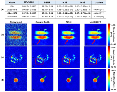
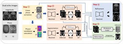
3845. Robust
Water-Fat Separation in Nasal MRI Using Hybrid Model and Data
Driven Network
D. Li, K. Sun, J. Zhang, D. Shen
School of Biomedical Engineering, & State Key Laboratory of Advanced Medical Materials and Devices, ShanghaiTech University, Shanghai, China
Impact: Our proposed hybrid separation framework
combines multi-task networks with a 3D physical model to
enhance the robustness of water/fat separation in
multi-contrast nasopharyngeal MRI images, significantly
expanding its clinical applicability.
|
 Back to Meeting Home
 Back to the Program-at-a-Glance
The International Society for Magnetic Resonance in Medicine is accredited by the Accreditation Council for Continuing Medical Education to provide continuing medical education for physicians.
 View
Presentation Video
View
Presentation Video