Digital Poster
Deep Learning for Image Enhancement: Part III
ISMRM & ISMRT Annual Meeting & Exhibition • 10-15 May 2025 • Honolulu, Hawai'i

Digital Poster
Deep Learning for Image Enhancement: Part III
 |
Computer Number: 33
3846. Consistently
denoising 3D MR images using 2D neural networks
M. Haas, M. Herbst
Bruker BioSpin GmbH & Co. KG, Ettlingen, Germany
Impact: The presented method enables consistent
high-dimensional denoising using 2-dimensional convolutional
networks. Thus, processing of large MRI datasets becomes
possible on standard workstations without the need of
expensive computer hardware or connection to a remote
server.
|
|
 |
Computer Number: 34
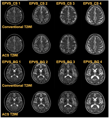
3847. AI-Accelerated
Brain MRI: 30% Faster Scans with Uncompromised Diagnostic
Accuracy for Aging Populations
W. Gu, C. Yang, Q. zhang, S. Cai, D. Wu, Y. Dai
Shanghai Punan Hospital of Pudong New Area, Shanghai, China
Impact: ACS dramatically reduces brain MRI scan time
while maintaining diagnostic accuracy. This offers a
practical, time-efficient alternative for routine
neuroimaging, particularly in elderly populations and
resource-limited settings, enhancing patient comfort and
clinical workflow without sacrificing diagnostic quality.
|
|
 |
Computer Number: 35
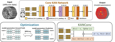
3848. DiffKAN:
Convolutional Kolmogorov-Arnold Networks for Improved Diffusion
MRI Microstructural Modeling
Y. Chen, Z. Li, Y. Wang, Z. Li, J. Zheng, H. Yang, M. Liu,
Q. Tian
Hangzhou Dianzi University, Hangzhou, China
Impact: DiffKAN’s efficient KAN-based architecture
offers a pathway to accurate diffusion MRI modeling
and analysis, significantly lowering computational burdens.
DiffKAN might transform the clinical adoption of diffusion
MRI, allowing for more widespread use in diagnostics by
providing more accurate microstructural mapping.
|
|
 |
Computer Number: 36
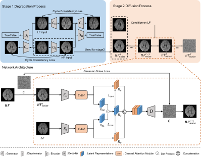
3849. Enhanced
Diffusion MRI of Infant Brain at 0.35 Tesla Using a
Self-Training Two-Stage Framework
Z. Chen, Y. Ding, H. Xu, L. Zhao, H. Zhang, D. Wu
Zhejiang University, Hangzhou, China
Impact: The proposed method improved the image quality
of DWI at low-field without collecting paired LF-HF data. It
may promote accurate diagnosis using low-field MRI for DWI.
|
|
 |
Computer Number: 37
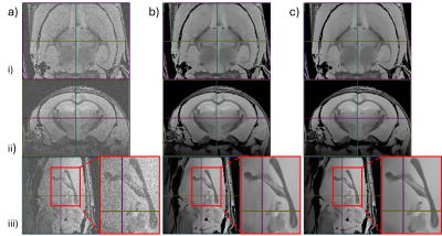
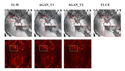
3850. Feasibility
study of Attention-Guided Pix2Pix GAN for the synthesizing of
enhanced cerebral vascular images in 7 Tesla MRI
J. Tan, Z. Zhen, Q. Wang, W. Chen, Z. Wang, W. Chen
Department of Medical Engineering, First Affiliated Hospital of Army Medical University, Chongqing, China
Impact: The model we proposed can generate high-quality
7T magnetic resonance cerebrovascular enhancement images,
which makes it possible to diagnose cerebral vessels without
the use of contrast agents.
|
|
 |
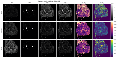
Computer Number: 38
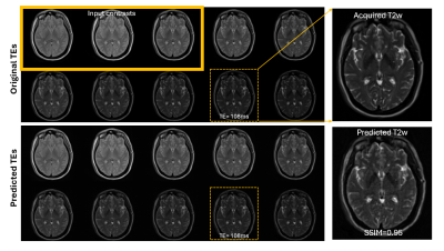
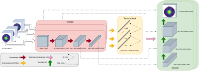
3851. Learning
temporal characteristics in multi-contrast MR images with
self-supervision: An application to accelerating quantitative T2
mapping
L. Umapathy, H. Pei, N. Ben-Eliezer, D. Sodickson, L. Feng
NYU Grossman School of Medicine, New York, United States
Impact: An understanding of underlying temporal
characteristics of tissues with vision transformers can help
with intelligent design of current multi-contrast data
acquisition schemes.
|
|
 |
Computer Number: 39
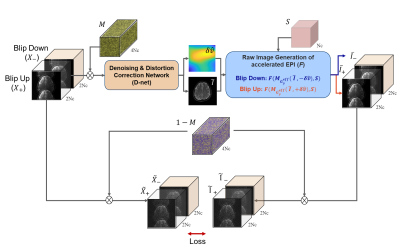
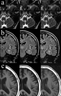
3852. Self-supervised
deep learning model for denioising and distortion correction in
accelerated echo planar imaging
J. Kim, B. Kang, H. Park, H. Seo
Korea Institute of Science and Technology (KIST), Seoul, Korea, Republic of
Impact: The proposed self-supervised approach achieves a
significant advancement by enabling simultaneous denoising
and distortion correction in accelerated EPI without ground
truth images, thereby enhancing image quality in accelerated
imaging.
|
|
 |
Computer Number: 40
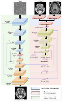
3853. MRI-Enhanced
Generative Model for Alzheimer's Disease: Converting 18F-FDG PET
to 18F-AV-45 PET
X. Fu, Y. Jin, H. Shao, H. Liu, Y. Zhang, N. Zhang, H.
Zheng, D. Liang, J. Liu, Z. Hu
Research Center for Medical AI, Shenzhen Institute of Advanced Technology, Chinese Academy of Sciences, Shenzhen, China
Impact:
This method significantly enhances diagnostic efficiency in Alzheimer's disease by enabling quick, cost-effective image generation, reducing reliance on expensive, short-lived tracers, and providing accessible support for clinicians, ultimately advancing multi-modal medical imaging practices in neurodegenerative diseases. |
|
 |
Computer Number: 41
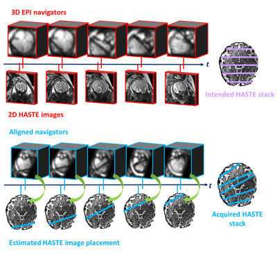
3854. 3D
fetal head pose estimation from MRI navigators with equivariant
networks
R. Muthukrishnan, B. Billot, B. Gagoski, M. Firenze, M.
Soldatelli, P. E. Grant, P. Golland
Massachusetts Institute of Technology, Cambridge, United States
Impact: Our method demonstrates the promise of fetal
head pose estimation and opensnew possibilities for dynamic
prescription of the imaging plane that tracks thefetal head
in real time.
|
|
 |
Computer Number: 42
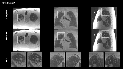
3855. Advanced
ZTE MR Lung Imaging: A Deep Learning Approach to Enhance SNR and
Reduce Artifacts
J. de Arcos, S. Mandava, M. Lebel, F. Wiesinger, C. Cretu,
P. Wielopolski, J. Hernández Tamames, P. Ciet
GE HealthCare, Chalfont Saint Giles, United Kingdom
Impact: The DL-ZTE model significantly enhances lung MRI
quality by reducing artifacts and improving SNR while
maintaining anatomical accuracy, facilitating more accurate
and reliable clinical assessments of parenchymal
abnormalities in ZTE lung imaging applications.
|
|
 |
Computer Number: 43
3856. Spatiotemporal
4D-UNet for Physics Consistent Super-Resolution and Denoising of
4DFlow-MRI
A. Ghazipour, A. Kazemi, A. Amini
University of Louisville, Louisville, United States
Impact: Our model can improve 4D-Flow MRI data that has
been hindered by noise, artifacts, and lower resolution.
As a result, hemodynamic parameters that are critical for
diagnosing various disease can be more accurately measured
|
|
 |
Computer Number: 44
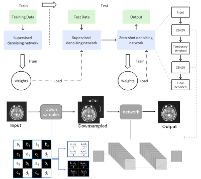
3857. A
Dual-Stage Denoising Method Based on Zero-Shot Learning for
Low-Field MRI
Y. Li, S. Liu, Y. Liu, M. Lyu
Shenzhen Technology University, Shenzhen, China
Impact: The framework of our proposed dual-stage
denoising method is plug-and-play for various existing
denoising models and generally enhances their performance.
|
|
 |
Computer Number: 45
3858. Specialized
Coil Informed Deep Learning for High SNR Carotid Imaging
L. Zeng, M. Lu, Y-C Hsu, M. Keushkerian, K-L Nguyen, K.
Johnson, M. Altbach, H. D. Morris, J. K. DeMarco, V.
Deshpande, D. Mitsouras, D. Saloner, S. McNally, S-E Kim, J.
Roberts, R. Hadley, G. Treiman, D. Parker, D. Li, Y. Xie
Cedars-Sinai Medical Center, Los Angeles , United States
Impact: With the use of our DL model, high SNR images
are achievable with standard head-neck coils, which may help
radiologists to be more confident and efficient in
evaluating carotid plaque characteristics.
|
|
 |
Computer Number: 46
3859. Simultaneous
reduction of noise and motion artifacts in brain MRI using deep
learning.
I. Muro, S. Shibukawa, K. Usui
AIC YAESU CLINIC, Tokyo, Japan
Impact: By 36,000 pairs of training data, we were able
to increase the accuracy of the learning process. The
advantage of this method is that it is post-processing and
can be used regardless of the equipment or imaging method.
|
|
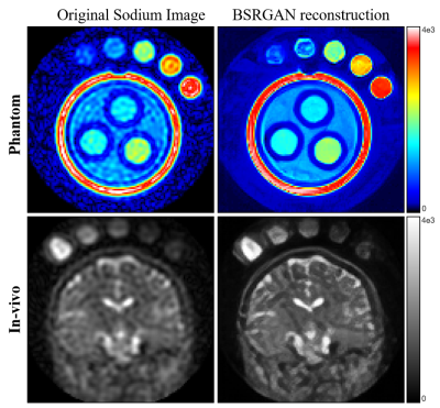 |
Computer Number: 47
3860. Enhanced
Sodium Imaging at 3T MRI Using BSRGAN: Achieving High SNR and
Spatial Resolution
S. Kim, S. Oh, S-Y Kim, J. Hwang, C. Y. Lim, Y. Sim, E. Kim,
E. S. Ko, W. K. Jeong, S. T. Kim, J. H. Lee
Department of Radiological Science, Daewon University College, Jecheon, Korea, Republic of
Impact: BSRGAN-based reconstruction of sodium images
achieved a 1.4-fold increase in signal-to-noise ratio and a
twofold improvement in spatial resolution at 3T,
significantly enhancing image quality and reducing
acquisition time.
|
 Back to Meeting Home
 Back to the Program-at-a-Glance
The International Society for Magnetic Resonance in Medicine is accredited by the Accreditation Council for Continuing Medical Education to provide continuing medical education for physicians.
 View
Presentation Video
View
Presentation Video