Digital Poster
AI for Image Segmentation: From Head to Toe
ISMRM & ISMRT Annual Meeting & Exhibition • 10-15 May 2025 • Honolulu, Hawai'i

Digital Poster
AI for Image Segmentation: From Head to Toe
 |
Computer Number: 49
1706. InvYNet:
an inverted Y shape network for prostate cancer segmentation
using prostate zones information
Y. Liu, Z. ZHU, X. Zhang, B. Zhang
The Affiliated Drum Tower Hospital of Nanjing University Medical School, Nanjing University, Nanjing, China
Impact: InvYNet is the first model to integrate
anatomical zones for prostate cancer segmentation, setting a
new standard in this field.
|
|
 |
Computer Number: 50
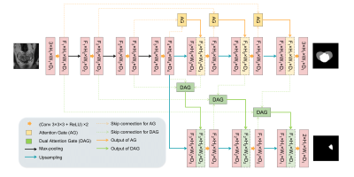
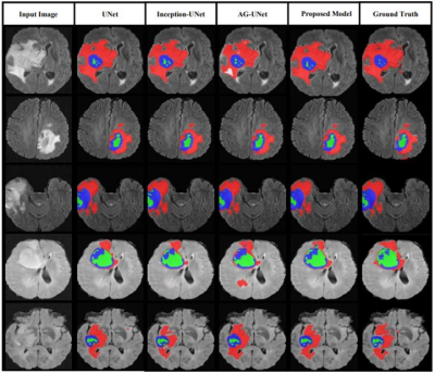
1707. Enhancing
Glioma Segmentation Accuracy Using Attention ResUNet
F. Moodi, F. Khodadadi Shoushtari, G. Valizadeh, D.
Mazinani, H. Mobarak Salari, H. Saligheh Rad
Quantitative MR Imaging and Spectroscopy Group (QMISG), Tehran University of Medical Sciences, Tehran, Iran-School of Medicine, Iran University of Medical Sciences, Tehran, Iran, Tehran, Iran (Islamic Republic of)
Impact: AResUNet demonstrates improved segmentation
performance for glioma brain tumors, offering insights that
may enhance diagnostic accuracy and treatment strategies in
clinical practice. This model's architecture showcases the
benefits of integrating attention mechanisms in deep
learning approaches for medical image analysis.
|
|
 |
Computer Number: 51
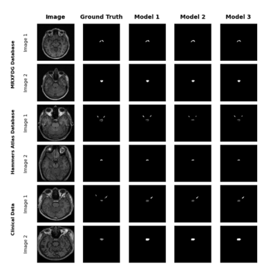
1708. Clinical
Evaluation of nn-UNet for Automated Segmentation of Pituitary
Gland and Optic Apparatus in Brain MRI: A Multi-Database
Approach
M. Yakubu, Q. Chen, A. Albusaidi, J. Shapey, A. King, A.
Hammers
King's College London, London, United Kingdom
Impact: This research has the potential to improve the
accuracy of MRI diagnostics for pituitary and sellar region
disorders. By addressing the challenges of model performance
on clinical data, it opens new avenues for optimizing deep
learning applications in medical imaging.
|
|
 |
Computer Number: 52
1709. Deep
Learning Based Tumor Segmentation on MRI of Prostate Cancer
Patient-Derived Xenografts in Mouse Models
S. Nayak, H. Salkever, E. Diaz, A. Sinha, N. Deveshwar, M.
Hess, M. Gibbons, S. Sahin, A. Rajagopal, P. Larson, R.
Sriram
University of California, San Francisco, San Francisco, United States
Impact:
This automated segmentation pipeline enhances efficiency in preclinical tumor studies, reducing manual effort and interuser variability. It provides a robust tool for evaluating treatment efficacy, potentially enabling broader use in diverse xenograft studies and informing translational research. |
|
 |
Computer Number: 53
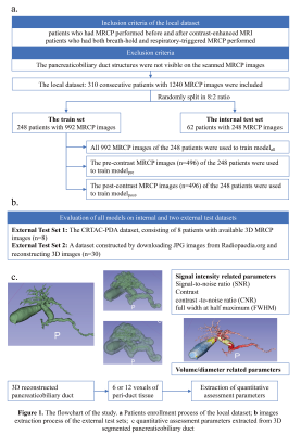
1710. Volumetric
Auto-Segmentation of the Pancreaticobiliary System for
Evaluating MRCP Image Quality: Efficacy Before and After
Contrast-Enhanced
z. zhou, C. Wang, S. Li, Z. Li
Huazhong University of science and technology, Tongji College, Tongji Hospital, wuhan, China
Impact: The automatic segmentation model provides a
robust tool for efficient 3D pancreaticobiliary
reconstruction, improving MRCP workflow. Future studies may
investigate optimizing MRCP quality based on patient factors
or clinical scenarios, and assess diagnostic value of
quantitative parameters extracted from segmentation.
|
|
 |
Computer Number: 54
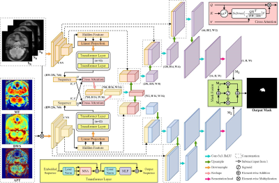
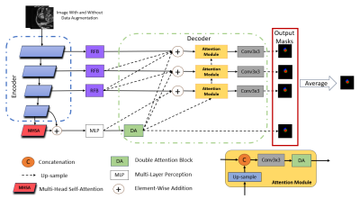
1711. CEST-Enhanced
Dual TransUNet for Precise Segmentation of Nasopharyngeal
Carcinoma
Z. Liyan, G. Cai, C. yingying, Y. Qian, C. Wei, L. Jianzhon,
L. Zhou
Department of Radiology, National Cancer Center/National Clinical Research Center for Cancer/Cancer Hospital & Shenzhen Hospital, Chinese Academy of Medical Sciences and Peking Union Medical College, 113 Baohe Avenue, 518116, Shenzhen, China, Shenzhen, China
Impact: The deep-learning model effectively utilized
CEST contrast for precise NPC segmentation, enhancing
radiotherapy planning by accurately targeting carcinoma and
preserving healthy tissue, while advancing CEST imaging's
role in clinical oncology.
|
|
 |
Computer Number: 55
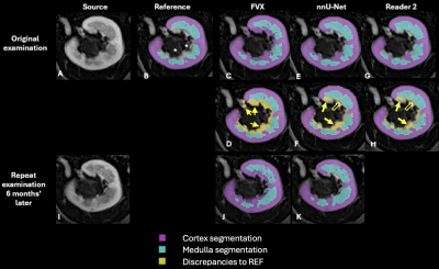
1712. Renal
corticomedullary segmentation from arterial phase volumetric
T1-weighted imaging: human or machine?
D. Liyanage, S. Kachel, L. McKenna, E. Hornsey, C.
Gillespie, B. Churilov, E. Ekinci, H. Rusinek, A. Mikheev,
R. Lim
Austin Health, Melbourne, Australia
Impact: MRI-derived renal corticomedullary segmentation
can be efficiently and reproducibly performed using
automated techniques with similar results to manual
segmentation. Such techniques have promise for assessment
and monitoring of chronic kidney disease and have potential
application for prognostication through multi-parametric
approaches.
|
|
 |
Computer Number: 56
1713. Cartilage
Auto-Segmentation of 3D T2* GRE Sequence in 7T High-Resolution
3D MRI
E. Hedayati, A. W. Kajabi, K. Knutsen, C. Steinberger, A.
Lamba, L. Tollefson, G. Metzger, R. LaPrade, J. Ellermann
University of Minnesota, Minneapolis, United States
Impact: This method accelerates cartilage segmentation
in 3D T2*-weighted MRI, reducing manual correction, speeding
ground truth creation, potentially supporting quantitative
analysis, and enhancing efficiency in cartilage assessment
for knee osteoarthritis.
|
|
 |
Computer Number: 57
1714. A
Shape Attentive Convolutional Neural Network for Improving the
Generalizability of CMR Image Segmentation
X. Wang, S. Lloyd, H. Gupta, L. Dell’Italia, T. Denney
Auburn University, Auburn, United States
Impact: Our network can be trained and validated on CMR
data from one site and can accurately segment CMR data from
other sites.
|
|
 |
Computer Number: 58
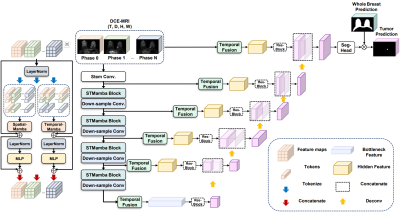
1715. Spatial-Temporal
Mamba Network for Accurate Breast Tumor Segmentation in DCE-MRI
H. Zhang, M. Wang, Y. Ren, J. Wen, W. Cui, B. Han, D. Luo,
Z. Liu, N. Zhang
Faculty of Robot Science and Engineering, Northeastern University, Shenyang, China
Impact:
Our results show that the proposed model can significantly improve tumor segmentation accuracy in DCE-MRI by utilizing both spatial and temporal features. This advancement holds promise for more accurate breast cancer diagnosis and better-informed treatment planning. |
|
 |
Computer Number: 59
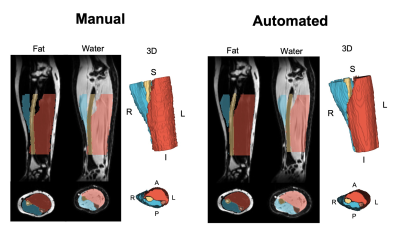
1716. Automated
MRI-Based Quantification of Forearm Muscle Health and
Associations with Hand Function
J. Fundaun, V. Oliva, S. Bédard, E. Wesselink, B. Lynn, A.
Pai S, D. Pfyffer, M. Kaptan, N. Berhe, J. Ratliff, S. Hu,
Z. Smith, T. Hastie, S. Mackey, M. McKay, J. Elliott, S.
Delp, G. Glover, A. Chaudhari, C. Law, A. Smith, K. Weber II
Stanford University, Palo Alto, United States
Impact: We developed an accurate, reliable
computer-vision model to automatically segment forearm
muscles, which will be made openly available. This method
can improve clinical assessment of forearm muscle health
leading to more efficient evaluation and management of
conditions affecting hand function.
|
|
 |
Computer Number: 60
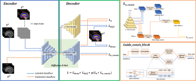
1717. DG-Net:A
Semi-Supervised Fetal Brain Segmentation Method Based on
Diffusion Model and Guided Consistency
K. Qi, C. Yan, D. Niu, B. Zhang, D. Liang, X. Long
Research Center for Medical AI, Shenzhen Institute of Advanced Technology, Chinese Academy of Sciences, Shenzhen 518055, China , Shenzhen, China
Impact: This study introduces a semi-supervised fetal
brain tissue segmentation method leveraging the diffusion
model and guided consistency. It achieves comparable
performance with fewer labeled samples, reducing manual
marking time and advancing fetal brain diagnosis.
|
|
 |
Computer Number: 61
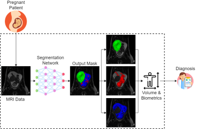
1718. Automated
Multi-Organ Segmentation in Fetal MRI
A. Lim, M. Wagner, B. Ertl-Wagner, L. Vidarsson, D. Sussman
Toronto Metropolitan University, Toronto, Canada
Impact: The Fetal MRI Segmentation Network (FetSegNet)
enables precise fetal body, amniotic fluid, and placenta
segmentation, enhancing clinical efficiency and supporting
more accurate pregnancy monitoring, paving the way for
improved maternal-fetal health diagnostics and a deeper
understanding of fetal development.
|
|
 |
Computer Number: 62
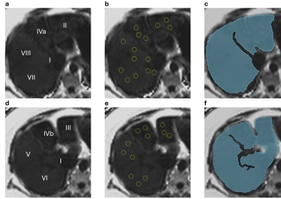
1719. Validation
of fully-automated whole liver segmentation for measurement of
hepatic fat fraction
N. Mahalingam, D. Bachu, C. Crabtree, K. Binzel, A. M.
Castillo, J. Volek, Y. Han, O. Simonetti
The Ohio State University, Columbus, United States
Impact:
Hepatic fat fraction from MRI can non-invasively stage the degree of hepatic steatosis for NAFLD evaluation. Automatic fat fraction measurement is more efficient than manual approaches, making it more suitable for clinical workflows. |
|
 |
Computer Number: 63
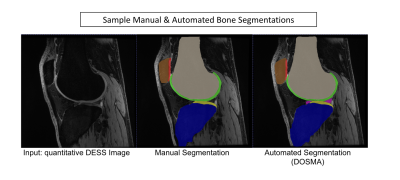
1720. Validation
of an Automated Open Source Pipeline for Comprehensive Knee MRI
Segmentation and Measurement of Quantitative Outcomes
F. Belibi, V. Sahani, Y. Vainberg, A. Goyal, A. Williams, C.
Chu, R. Pedersen, B. Haddock, A. Chaudhari, F. Kogan, A.
Gatti
Stanford University, Stanford, United States
Impact: Automated segmentation and analysis of bone and
cartilage have the potential to greatly improve the
translation potential of quantitative MSK MRI biomarkers.
|
|
 |
Computer Number: 64
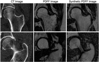
1721. Data
Augmentation with Generative Deep Learning for Automatic Bone
Segmentation from Fat Fraction MRI
N. Dwork, P. Elangovan, D. Connor, A. McManus, R. Krug, G.
Kazakia, C. Jankowski, J. Carballido-Gamio
University of Colorado Anschutz, Aurora, United States
Impact: With an automatic bone segmentation algorithm
from fat fraction MR images, future work will conduct a
thorough investigation into how bone marrow adiposity
affects bone fragility.
|
 Back to Meeting Home
 Back to the Program-at-a-Glance
The International Society for Magnetic Resonance in Medicine is accredited by the Accreditation Council for Continuing Medical Education to provide continuing medical education for physicians.
 View
Presentation Video
View
Presentation Video