Digital Poster
Segmentation
ISMRM & ISMRT Annual Meeting & Exhibition • 10-15 May 2025 • Honolulu, Hawai'i

 |
Computer Number: 33
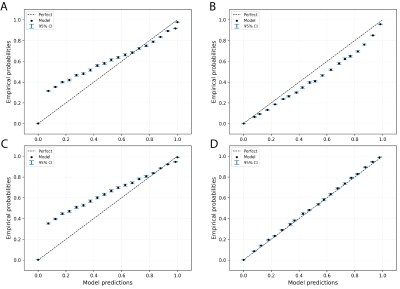
4301. Applying
Venn-Abers Predictors to Calibrate White Matter Hyperintensity
Segmentations from Brain Images
K. Landheer, K. Landheer, B. Geraghty, J. Herman, N.
Parikshak, M. Goubran, J. Marchini
Regeneron Genetics Center, Tarrytown, United States
Impact: We demonstrated that Inductive Venn-Abers
Predictors can be used to reliably calibrate a deep-learning
segmentation tool, which improved model performance,
calibration, uncertainty estimates, and aids in the
interpretability of the resulting segmentation maps
|
|
 |
Computer Number: 34
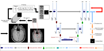
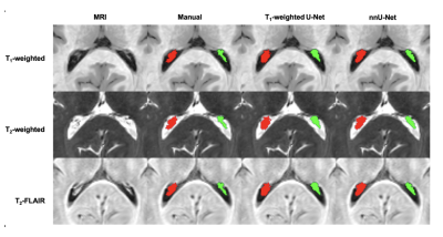
4302. Improving
Substantia Nigra Segmentation Across Different
Neuromelanin-Sensitive MRI Sequences Using Domain Generalization
Techniques
O. Welsh, K. Hett, A. Bosman, D. Claassen, P. Trujillo
Vanderbilt University Medical Center, Nashville, United States
Impact: Applying data augmentation techniques
significantly enhances automated substantia nigra
segmentation in neuromelanin-sensitive MRI, advancing the
development of robust, clinically reliable models adaptable
to various imaging methods and neurodegenerative conditions.
|
|
 |
Computer Number: 35
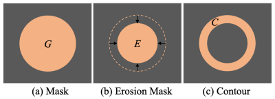
4303. Improving
medical image segmentation using contour-weighted loss
Z. Huang, N. Jiang, Y. Sui
National Institute of Health Data Science, Peking University, Beijing, China
Impact: We developed a contour-weighted loss function to
address the problem of data imbalance in medical image
segmentation. Our approach is model-independent, allowing it
to integrate seamlessly with any segmentation network,
thereby improving segmentation performance across different
models.
|
|
 |
Computer Number: 36
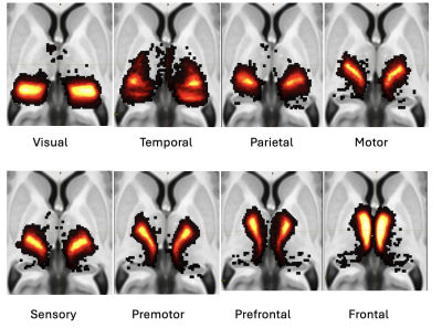
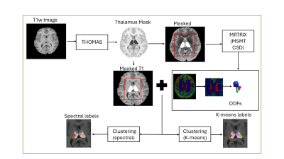
4304. Revisiting
the role of structural connectivity in thalamic nuclei
segmentation
D. Nguyen, V. Kumar, D. Patterson, M. Saranathan
UMass Chan Medical School, Worcester, United States
Impact: These results advance our understanding of
thalamic connectivity, potentially guiding targeted clinical
interventions and personalized therapies for neurological
conditions. This study enables future research on
connectivity-driven parcellation techniques, raising
questions about refining segmentation for enhanced
anatomical accuracy.
|
|
 |
Computer Number: 37
4305. Multi-contrast
deep-learning segmentation of the choroid plexus using
self-configuring nnU-Net
K. Bagai, A. Song, M. Leguizamon, A. Dubois, C. McKnight, C.
Considine, P. Trujillo, D. Claassen, M. Donahue, K. Hett
Vanderbilt University Medical Center, Nashville, United States
Impact: This study evaluates multi-contrast MRIs as
inputs to a self-configuring deep learning framework to
provide a new tool for segmentation of the choroid plexus,
which has gained much recent interest as the most proximal
structure in the neurofluid circuit.
|
|
 |
Computer Number: 38
4306. Validation
of Deep Learning based Tissue Segmentation for Efficient and
Robust Quantitation of Background Parenchymal Enhancement on
Breast MRI
Y-T Kuo, A. Kazerouni, V. Park, W. Surento, S.
Sujichantararat, D. Hippe, H. Rahbar, S. Partridge
University of Washington, Seattle, United States
Impact: Application of deep learning for segmentation of
fibroglandular tissue on breast MRI can improve the
robustness and reliability of quantitative imaging
biomarkers, with the potential to improve risk
stratification and clinical decision-making for high-risk
breast cancer screening.
|
|
 |
Computer Number: 39
4307. Fast
and Efficient Diffusion-based Thalamic Segmentation Using
Spectral Clustering
D. Das, C. Iglehart, A. Bilgin, M. Saranathan
University of Arizona, Tucson, United States
Impact: Fast and accurate subthalamic segmentation can
enable more accurate thalamic studies and interventions,
thereby improving both our understanding of brain pathology
and patient outcomes in various neurological conditions
|
|
 |
Computer Number: 40
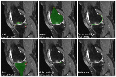
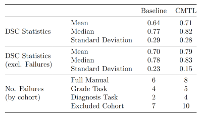
4308. Fully
Automatic Segmentation of Knee Joint Anatomy and Lesions using
Clinical MR Images
A. Yu, M. Yang, S. Tosun, R. Lartey, K. Nakamura, N. Subhas,
C. Winalski, X. Li
Cleveland Clinic, Cleveland, United States
Impact: We provide an efficient and consistent solution
for the segmentation of knee joint anatomy and lesions,
enabling large-scale downstream analyses without incurring
large costs for manual annotations.
|
|
 |
Computer Number: 41
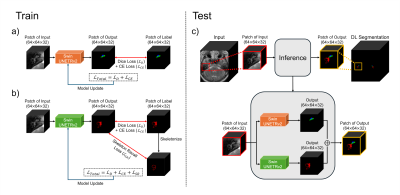
4309. Cross-Modal
Transfer Learning Enables Clinically Useful Segmentation of
Pediatric Brain Tumors using Diffusion Weighted Imaging
T. Mulvany, D. Griffiths-King, H. Rose, J. Apps, A. Peet, J.
Novak
Aston University, Birmingham, United Kingdom
Impact: Establishes benefits of leveraging large non-DWI
public datasets, to improve automated DWI segmentation
models, essential for native pediatric brain tumour
analysis. This eliminates error arising from image
co-registration, streamlines clinical workflows and limits
the impact of missing imaging modalities.
|
|
 |
Computer Number: 42
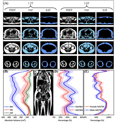
4310. Reproducibility
of automatic adipose tissue segmentation using PDFF images
between 1.5T and 3.0T MR
C. Cheng, J. Gong, H. Peng, Q. Wan, X. Liu, H. Zheng, C. Zou
Shenzhen institutes of advanced technology, Chinese Academy of Sciences, Shenzhen, China
Impact: These findings could improve adipose tissue
assessment in diverse clinical MR settings. This enhancement
would enable large cohort studies to better identify
obesity-related health risks using multicenter datasets,
thus facilitating a more effective approach to obesity
management.
|
|
 |
Computer Number: 43
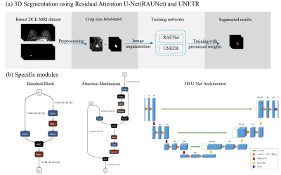
4311. Deep
Learning-enabled Fully Automated 3D DCE-MRI Segmentation for
Breast Cancer Lesion
R. Zhang, K. Wang, S. Huang, J. Xie, S. Wang, M. Xu
The First Affiliated Hospital of Zhejiang Chinese Medical University (Zhejiang Provincial Hospital of Chinese Medicine), hangzhou, China
Impact: By constructing and training an efficient deep
learning model to achieve high-precision segmentation of
breast cancer lesions, it provides a powerful auxiliary tool
for clinical diagnosis, treatment planning and prognosis
analysis.
|
|
 |
Computer Number: 44
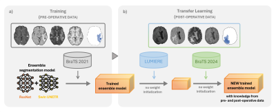
4312. Leveraging
transfer learning for post-operative brain tumor segmentation
across MRI datasets
C. Passarinho, O. Lally, A. Matoso, M. Loureiro, J. Moreira,
P. Figueiredo, R. Nunes
Instituto Superior Técnico, Universidade de Lisboa, Lisbon, Portugal
Impact: This work addresses the hurdles of automated
post-operative tumor segmentation by demonstrating that
transfer learning from pre-operative models can improve
post-treatment segmentation. The importance of large
annotated datasets and the effects of catastrophic
forgetting and model knowledge retention are highlighted.
|
|
 |
Computer Number: 45
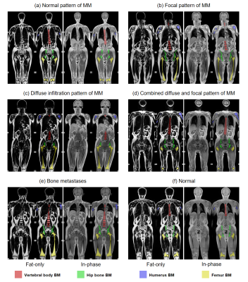
4313. Segmentation
model quantifying bone marrow fat on whole-body Dixon MRI
reveals association between vertebral fat and diabetes
S. Huang, Q. Wang, F. Cong, J. Zhu, Z. Jin, H. Xue
Peking Union Medical College Hospital, Beijing, China
Impact: Our novel three-dimensional nnU-Net model for
automated assessment of whole-body bone marrow fat sheds new
light on the link between bone marrow adiposity and
diabetes.
|
|
 |
Computer Number: 46
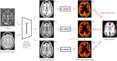
4314. Enhancing
Reliability of MRI-based Brain Morphometry by Synthetic MPRAGE
Generation
T. Blattner, R. McKinley, R. Wiest, C. Rummel, M. Capiglioni
Institute for Diagnostic and Interventional Neuroradiology, University of Bern, Bern, Switzerland
Impact: We created a contrast-invariant segmentation
tool that improves brain morphometry accuracy across
variable MRI settings, enabling more reliable monitoring of
neurodegenerative disease progression. This tool improves
assessment accuracy across longitudinal and multi-parameter
MRI acquisitions common in clinical practice.
|
|
 |
Computer Number: 47
4315. Deep
Learning-Based Topology-Preserving Inner Ear Subregion
Segmentation in MRI
W. Kim, D. Bak, Y. Kang, H-J Lee, Y. Nam
Hankuk University of Foreign Studies, Yongin, Korea, Republic of
Impact: The proposed inner ear subregion segmentation
method may aid in diagnosing and planning treatment for
auditory-related conditions, such as Meniere’s disease, by
enabling automatic quantification of contrast enhancement
for each inner ear region.
|
|
 |
Computer Number: 48
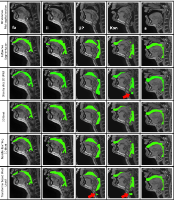
4316. A
comparative study on state-of-the-art deep learning based vocal
tract segmentation methods in volumetric sustained speech MRI
S. Erattakulangara, S. Gerard, D. Meyer, K. Kelat, K.
Burnham, R. Balbi, S. Lingala
The University of Iowa, iowa city, United States
Impact: This study informs researchers about various
state-of-the-art segmentation methods for upper airway MRI.
It emphasizes the strengths and weaknesses of each method
and identifies which methods work efficiently under specific
conditions.
|
 Back to Meeting Home
 Back to the Program-at-a-Glance
The International Society for Magnetic Resonance in Medicine is accredited by the Accreditation Council for Continuing Medical Education to provide continuing medical education for physicians.
 View
Presentation Video
View
Presentation Video