Digital Poster
Everything & Every MRI in Hepatobiliary & GI
ISMRM & ISMRT Annual Meeting & Exhibition • 10-15 May 2025 • Honolulu, Hawai'i

Digital Poster
Everything & Every MRI in Hepatobiliary & GI
 |
Computer Number: 97
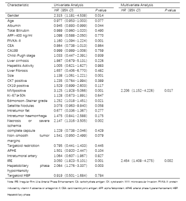
2821. Pathological
Features of Hepatocellular Carcinoma with Irregular Rim-Like
Arterial Phase Enhancement and its Prognosis Analysis
Z. Yan, X. Zhang, L. Xu, X. Zhao
Nantong University, Nantong Third People’s Hospital, Affiliated Nantong Hospital 3 of Nantong University, Nantong, China
Impact: HCC with irregular rim-like enhancement in the
arterial phase may serve as a non-invasive biomarker for
predicting poor prognosis.
|
|
 |
Computer Number: 98
2822. Washout
on Delayed Post Contrast High Resolution Spoiled Gradient Echo
MRI is a Sign of Residual Rectal Cancer after Neoadjuvant
Therapy
M. U. Nisar, S. Raichandani, A. Pratapneni, P. Ghanouni, E.
Pollom, V. Sheth
Department of Radiology, Stanford University, Palo Alto, United States
Impact: Gadolinium washout may be useful in conjunction
with T2-weighted and diffusion-weighted images for accurate
assessment of rectal cancer treatment response.
|
|
 |
Computer Number: 99
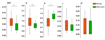
2823. Non-invasive
prediction of KRAS mutation in rectal cancer using hybrid
intravoxel incoherent motion and diffusion kurtosis model
J. YUAN, M. Liu, W. Tan
Shuguang Hospital Affiliated to Shanghai University of Traditional Chinese Medicine, Shanghai, China
Impact: The hybrid IVIM-DKI model, especially the K
value, demonstrates potential as a non-invasive imaging
biomarker for KRAS mutation status in rectal cancer. This
approach could guide treatment decisions and evaluate
prognosis without invasive biopsies, potentially improving
patient care and outcomes in rectal cancer management.
|
|
 |
Computer Number: 100
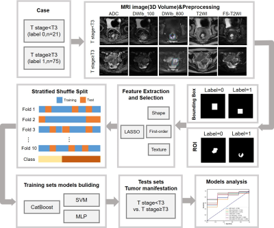
2824. Improve
Model Performance Rectal Cancer Staging by Radiomics Analysis of
Diffusion-weighted Imaging and T2-weighted Imaging
Y-C Chen, C-F Hsieh, S-C Lin, C-C Chang, C-J Juan, Y-J Liu,
H-H Peng
National Tsing Hua University, Hsinchu, Taiwan
Impact: Compared to extracting features from DWI or T2WI
solely, radiomics analysis of combinations of DWI_b800+T2WI
or DWI_b800+FS-T2WI exhibited better AUC and TNR in
identifying rectal cancer patients ≥T3. A bounding-box was
the best strategy for determining the analyzed region.
|
|
 |
Computer Number: 101
2825. Virtual
magnetic resonance elastography and fractional order calculus
model for assessing proliferation status in rectal carcinoma
N. Meng, Y. Cui, X. Wang, X. Chen, J. Guo, L. Xie, M. Wang
Henan Provincial People’s Hospital & Zhengzhou University People’s Hospital, Zhengzhou, China
Impact: The vMRE, FROC, and DWI can effectively
differentiate between high- and low-proliferation rectal
carcinoma. and a composite diagnostic tool consisting of µMRE,
D, and β may serve as a promising biomarker to help make
optimal clinical decisions.
|
|
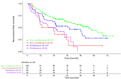 |
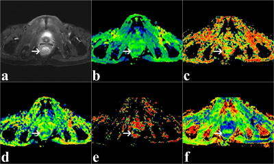
Computer Number: 102
2826. Imaging
features and prognosis after curative resection of proliferative
hepatocellular carcinoma with LR-4/5 and LR-M
Y. Xie, Z. Liu, X. Zhang, X. Zhao
Nantong University, Nantong Third People’s Hospital, Affiliated Nantong Hospital 3 of Nantong University, Nantong, China
Impact: Combining LI-RADS classification and
proliferation helps to stratify the risk of recurrence after
curative resection in patients with solitary HCC.
|
|
 |
Computer Number: 103
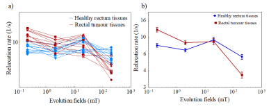
2827. In-vivo
detection of rectal cancer at low field using field cycling
imaging
A. Alamri, N. Senn, L. Samuel, R. Mitchell-Hay, D. Lurie, G.
Ramsay, L. Broche
University of Aberdeen, Aberdeen, United Kingdom
Impact: This FFC-NMRD technique could complement
existing imaging modalities by addressing limitations in
detecting post-therapy changes in rectal cancer, enabling
non-invasive diagnostics. Also, it holds potential to
improve personalized treatment, guiding more effective
therapies and enhancing patient outcomes.
|
|
|
Computer Number: 104
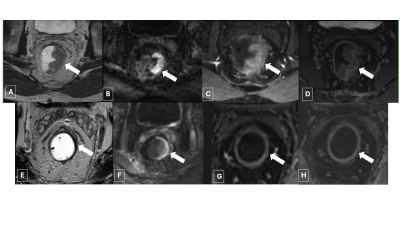
2828. FOCUS-MUSE
DWI in Primary Rectal Cancer: Comparison with FOCUS, MUSE, and
Single-shot EPI DWI
W. Feng, L. Zhu, K. Wang, J. Dai, Q. Ma, H. Shen, F. Yuan,
H. Zhang
Ruijin Hospital, Shanghai Jiao Tong University of Medicine, China, Shanghai, China
Impact: Our study showed FOCUS-MUSE provided improved
image quality of DWI for primary RC T staging. The
application of FOCUS-MUSE DWI may be beneficial for
evaluating and managing RC.
|
||
 |
Computer Number: 105
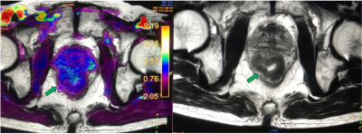
2829. T1-Mapping
with High-Spatial-Resolution T2-Weighted Imaging Differentiates
Mucinous from Nonmucinous Adenocarcinoma in Rectal Cancer
Y. Liu, Z. Wen, Y. Chen, S. Yu, Y. Ma, X. Yang, Y. Wu
The First Affiliated Hospital, Sun Yat-sen University, Guangzhou, China
Impact: This study suggests combining HR-T2WI and
T1-mapping as a non-invasive method for diagnosing MC in
rectal cancer, potentially reducing false-negative biopsies.
Future research can explore its role in treatment response
prediction and outcome assessment.
|
|
 |
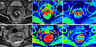
Computer Number: 106
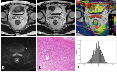
2830. The
evaluation of MR staging of rectal cancer by using amide proton
transfer weighted (APTw) imaging
Y. Gao, J. Liu, J. Lian, B. Pan, J. Qiu
Peking University First Hospital, Beijing, China
Impact: APTw imaging provides information on rectal
cancer staging.
|
|
 |
Computer Number: 107
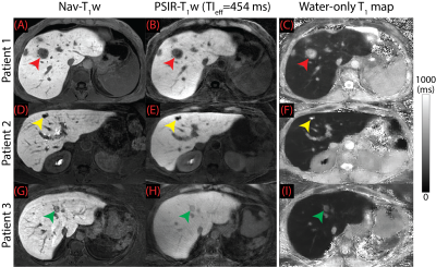
2831. Improved
Characterization of Liver Lesions with Hepatobiliary Phase
Free-Breathing, Phase Sensitive Inversion Recovery MRI
Y. Muslu, J. Heidenreich, J-P Grunz, T. Cashen, S. Mandava,
A. Pirasteh, D. Hernando, S. Reeder
University of Wisconsin-Madison, Madison, United States
Impact: GA-enhanced PSIR-T1w
MRI improves T1 contrast
and offers variable T1w
imaging in a single acquisition, enabling both detection and
characterization of lesions with a single T1w
image acquisition.
|
|
 |
Computer Number: 108
2832. Efficient
Assessment of CR to Neoadjuvant Therapy in Locally Advanced
Rectal Cancer using Amide Proton Transfer Weighted MRI: a
Diagnostic Study
L. Zhang, Z. Jin, P. Sun, Y. Liu, X. Li
Union Hospital, Tongji Medical College, Huazhong University of Science and Technology, Wuhan, China
Impact: Our study revealed the clinical significance of
APTw MRI as a promising imaging modality for assessing CR to
NAT in LARC. APTw MRI can provide unique insights into tumor
metabolism and microenvironment, surpassing the capabilities
of traditional MRI.
|
|
 |
Computer Number: 109
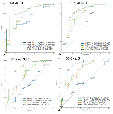
2833. Assessing
Liver Fibrosis with Gd-EOB-DTPA-enhanced B1
Inhomogeneity-corrected VFA T1 mapping: Comparison with US
Shear-wave Elastography
Y. Li, C. Fu
Department of Radiology,Zhongshan Hospital of Fudan University, Shang hai, China
Impact: It is essential to apply a noninvasive and
accurate method for predicting liver fibrosis stages. Liver
stiffness measurement using US shear-wave elastography is
superior to T1 relaxation times obtained from
Gd-EOB-DTPA-enhanced T1 mapping for staging liver fibrosis.
|
|
 |
Computer Number: 110
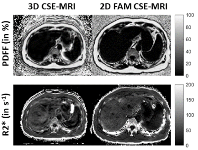
2834. Single-Site
Experience of Liver Fat and Iron Quantitation with a 2D Flip
Angle Modulated CSE-MRI Sequence in the Clinical Setting
E. Milshteyn, A. Guidon, J. Tang, S. Reeder, D. Hernando, N.
Nakrour, M. Harisinghani, A. Mojtahed
GE HealthCare, Boston, United States
Impact: With its free-breathing nature and reproducible
PDFF and R2* values compared to gold standard 3D CSE-MRI, 2D
FAM CSE-MRI shows promise as a routine tool in clinical
liver fat and iron quantitation.
|
|
 |
Computer Number: 111
2835. Application
Value of MR Elastography in the Identification of Benign and
Malignant Liver Lesions and Prognostic Prediction of Malignant
Tumors
X. Huang, W. Ma, Y. Sun, J. Wang, Z. Qin, W. Tan, J. Yuan
Shuguang Hospital Affiliated to Shanghai University of Traditional Chinese Medicine, Shanghai, China
Impact: MRE demonstrates potential as a non-invasive
imaging biomarker for distinguishing benign from malignant
liver lesions and assessing treatment response in malignant
tumors. This could facilitate more informed clinical
decisions and improve patient outcomes.
|
 Back to Meeting Home
 Back to the Program-at-a-Glance
The International Society for Magnetic Resonance in Medicine is accredited by the Accreditation Council for Continuing Medical Education to provide continuing medical education for physicians.
 View
Presentation Video
View
Presentation Video