Digital Poster
Promising Lung MR Applications
ISMRM & ISMRT Annual Meeting & Exhibition • 10-15 May 2025 • Honolulu, Hawai'i

 |
Computer Number: 49
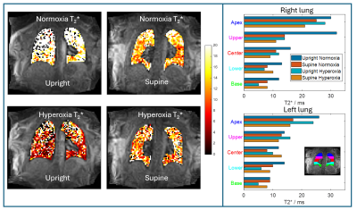
1865. Investigating
the Effect of Gravity and Oxygen Signal Enhancement on Lung T2*
with Upright 0.5T MR
Z. Peggs, O. Mougin, A. Harrison, S. Needleman, N. Blockley,
M. Kim, G. Pavlovskaya, T. Meersmann, S. Francis, G. Parker,
P. Gowland, R. Sobhan
University of Nottingham, Nottingham, United Kingdom
Impact: Oxygen-enhanced
lung MRI using a 0.5T upright scanner facilitates comparison
of parametric maps and ventilation at seated vs supine
postures. This can provide crucial knowledge for clinicians
and researchers in understanding and characterising lung
physiology and function.
|
|
 |
Computer Number: 50
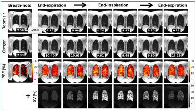
1866. Respiratory-resolved
lung oxygen and compliance mapping at 0.55T in patients with
lymphangioleiomyomatosis
J. Plummer, P. Daudé, R. Ramasawmy, A. Javed, A.
Tsakirellis, J. Moss, A. Campbell-Washburn
National Institutes of Health, Bethesda, United States
Impact: Respiratory-resolved oxygen-enhanced MRI offers
a temporal view of pulmonary oxygen perfusion, complemented
by additional insight into regional alveolar compliance from
specific ventilation (SV) images calculated from the same
data. This four-dimensional, patient-friendly approach
enables comprehensive assessment of lung function
abnormalities.
|
|
 |
Computer Number: 51
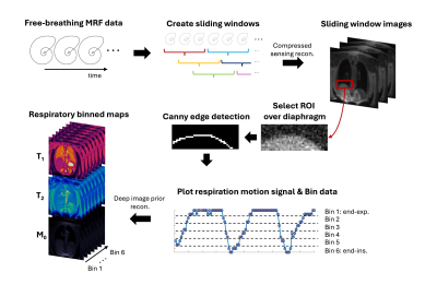
1867. Free-Breathing
Lung MR Fingerprinting at 0.55T with Retrospective Respiratory
Motion Binning
Z. Liu, N. Seiberlich, J. Hamilton
University of Michigan, Ann Arbor, United States
Impact: This work demonstrates feasibility of
free-breathing lung MR Fingerprinting for respiratory
motion-resolved 2D T1,
T2 and
M0 mapping
at 0.55T in healthy subjects, which may have potential
clinical applications to pulmonary conditions, such as
emphysema, COPD, and interstitial lung diseases.
|
|
 |
Computer Number: 52
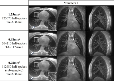
1868. Free-running
4D lung imaging at 0.55T using UTE-bSSFP with intra-bin
correction and inter-bin compensation of respiratory motion
A. Mackowiak, S. Rapacchi, G. Fahrni, C. Pozzessere, J-B
Ledoux, M. Stuber, D. Rotzinger, C. Roy
Centre Hospitalier Universitaire Vaudois and University of Lausanne, Lausanne, Switzerland
Impact: This study further validates low-field UTE lung
imaging, achieving sub-millimetric 4D resolution without
significant loss of pulmonary vessel sharpness or apparent
SNR when accelerating two-fold, thanks to a powerful
respiratory-resolved reconstruction, suggesting strengthened
clinical translation.
|
|
 |
Computer Number: 53
1869. 0.55T
Low-field MRI spirometry
C. Valle, J. Retamal, R. Salas, M. Andia, C. Besa
Millennium Institute for Intelligent Healthcare Engineering (iHealth), Santiago, Chile
Impact: This preliminary study using low-field MRI
spirometry reveals potential differences in lung function
between active and sedentary individuals. The findings
suggest that low-field MRI could serve as a viable, detailed
regional lung assessment, potentially benefiting management
of chronic lung pathologies.
|
|
 |
Computer Number: 54
1870. Free-breathing
PREFUL-MRI of the lungs at 0.55T detects functional alterations
in patients with lung lesions
J. Liu, W. Li, X. Wang, J. Zhu, X. Wang, R. GRIMM, J. Qiu
Peking University First Hospital, Beijing, China
Impact: This exploratory study demonstrates the
potential of free-breathing PREFUL-MRI at 0.55T in
identifying functional abnormalities in patients with
pulmonary lesions, suggesting its utility as a follow-up
tool.
|
|
 |
Computer Number: 55
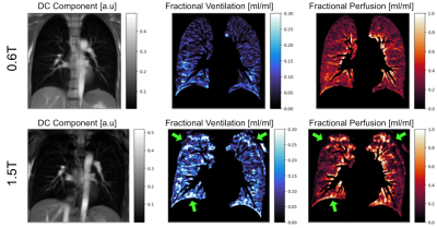
1871. Free-Breathing
Functional Lung Imaging at 0.6T compared to 1.5T
E. Ilicak, E. Ercan, Y. Dong, M. Staring, A. Webb, M. van
Osch, P. Börnert, M. Nagtegaal
Division of Image Processing , Leiden, Netherlands
Impact: We investigate the usefulness of 0.6T MRI for
free-breathing functional lung imaging. Our findings
demonstrate improved image quality compared to 1.5T, with
improved tissue-background contrast and homogeneity of
functional maps, underscoring the system's robustness and
potential for non-invasive pulmonary imaging.
|
|
 |
Computer Number: 56
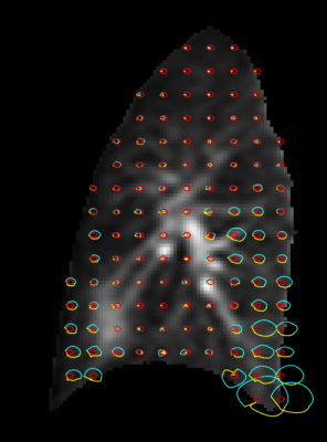
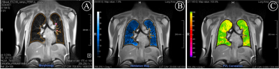
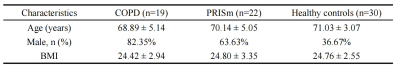
1872. Four-dimensional
dynamic ultrashort echo time MRI for functional imaging in
chronic lung diseases: A preliminary study
Z. Zhang, Z. Ding, J. Li, Y. Xia, Z. Wu, H. She, M. Xu, Y.
P. Du, L. Fan
Department of Radiology, Second Affiliated Hospital of Naval Medical University, Shanghai, China
Impact: The pulmonary dynamic UTE MRI allowed for
exhibiting ventilation inhomogeneity within free breathing
in patients with COPD and PRISm.
|
|
 |
Computer Number: 57
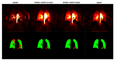
1873. SPGRE-to-bSSFP
Sequence-Mapping with U-Net and GAN for Data Homogenization
improves Comparability of PREFUL MRI
A. Voskrebenzev, J. Hahn, M. Zubke, F. Klimeš, M. Wernz, R.
Müller, F. Wacker, J. Vogel-Claussen
Hannover Medical School, Hannover, Germany
Impact: As sequence homogenization is limited by vendor
standards and hardware-limits the demonstrated
sequence-mapping approach via deep learning is viable
alternative. It could be specifically used to decrease the
variability of perfusion-weighted maps acquired with bSSFP
and SPGRE in multicenter settings.
|
|
 |
Computer Number: 58
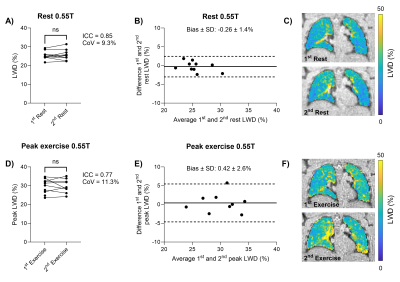
1874. Repeatability
and cross field strength reproducibility of lung water MRI at
rest and exercise stress
F. Seemann, N. Castor, A. Javed, R. Ramasawmy, J. Plummer,
G. Weissman, E. Morgan, A. Campbell-Washburn
National Heart, Lung, and Blood Institute, National Institutes of Health, Bethesda, United States
Impact: Dynamic lung water MRI during exercise stress
may have clinical utility in heart failure. Understanding
this method’s repeatability and performance across different
magnetic field strengths accelerates the clinical adoption
of this tool.
|
|
 |
Computer Number: 59
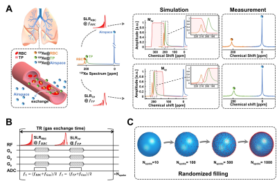
1875. Direct
imaging of pulmonary gas exchange in vivo with hyperpolarized
xenon MRI

H. Li, H. Li, M. Zhang, X. Liu, Y. Zheng, Y. Fang, Y.
Han, X. Zhou
State Key Laboratory of Magnetic Resonance and Atomic and Molecular Physics, National Center for Magnetic Resonance in Wuhan, Innovation Academy for Precision Measurement Science and Technology, Chinese Academy of Sciences - Wuhan National Laboratory for Optoelectronics , Wuhan , China
Impact: The
proposed gas exchange MRI method improves diagnosis of lung
diseases by accurately assessing gas exchange function in
the lungs.
|
|
 |
Computer Number: 60
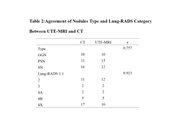
1876. Value of
ultrashort echo time MRI in pulmonary nodules detection and
Lung-RADS grading
S. Liu, X. Wang, Y. Wang, Y. Cui, N. Meng, W. Wei, Y. Bai,
Y. Shen, X. Zhang, T. Benkert, M. Wang
Department of Radiology, Xinxiang Medical University & Henan Provincial People’s Hospital, Zheng zhou, China
Impact: UTE now rivals the ‘gold standard’ chest CT scan
in the detection rate of pulmonary nodules and the ability
to clearly display their morphological characteristics;
it also shows a high degree of consistency in the Lung-RADS
grading assessment.
|
|
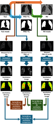 |
Computer Number: 61
1877. Impact
of inflation level on breath-hold 1H MRI surrogates of regional
lung ventilation.
W. Clark, J. Astley, A. Biancardi, L. Saunders, P. Hughes,
J. Wild, B. Tahir
University of Sheffield, Sheffield, United Kingdom
Impact:
This study demonstrates that breath-hold 1H-MRI ventilation surrogates vary significantly with inflation level. The strong agreement between measures derived from TLC-RV and FRC-RV suggests these combinations provide similar information, while other pairs may capture complementary aspects of regional ventilation distribution. |
|
 |
Computer Number: 62
1878. Ultrashort
echo-time (UTE) functional lung imaging for fractional
ventilation quantification: breath-hold vs. free-breathing
H. Liang, R. Zhang, P-Y Wu
GE HealthCare MR Research, Beijing, China
Impact: The proposed free-breathing UTE method is
straightforward to implement in clinical practice, and can
provide satisfactory quantitative FV maps in subjects who
fail to maintain breath-hold, improving the success rate of
clinical UTE functional lung imaging.
|
|
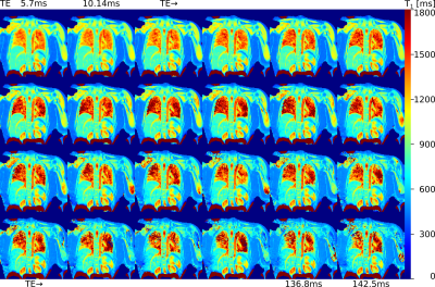 |
Computer Number: 63
1879. TE-dependent
observed lung T1 measured using inversion recovery radial turbo
spin echo
S. Triphan, K. Zhang, H-U Kauczor, M. Wielpütz
University Hospital Heidelberg, Heidelberg, Germany
Impact: Observed T1 in the lungs shows a dependence on
TE in gradient echo sequences due to different T2* and T1 in
tissue compartments. Here, we show TE-dependence due to T2
weighting and thus compartments also differ in T2.
|
|
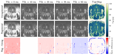 |
Computer Number: 64
1880. Feasibility
of T1rho Mapping with On-Scanner Non-rigid Image Registration
for Lung Imaging
R. Klaar, I. Benlala, K. Narceau, P. Gut, V. de Villedon de
Naide, T. Génisson, K. He, T. Richard, G. Dournes, M. Stuber,
J. Dinkel, A. Bustin
LMU University Hospital, LMU Munich, Munich, Germany, Munich, Germany
Impact: The acquisition of T1ρ-maps for lungs could
provide valuable additional information in the
identification and classification of inflamed and fibrotic
tissue regions present in diseases such as radiation-induced
pneumonitis or interstitial lung disease.
|
 Back to Meeting Home
 Back to the Program-at-a-Glance
The International Society for Magnetic Resonance in Medicine is accredited by the Accreditation Council for Continuing Medical Education to provide continuing medical education for physicians.
 View
Presentation Video
View
Presentation Video