Digital Poster
X-Nuclei
ISMRM & ISMRT Annual Meeting & Exhibition • 10-15 May 2025 • Honolulu, Hawai'i

 |
Computer Number: 49
3072. Application
of the power independent of number of slices presaturated
ultrashort echo time (PINS-UTE) sequence to sodium MRI
J. Reich, E. MacMillan, R. Feldman
University of British Columbia, Kelowna, Canada
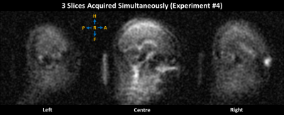
Impact: We translate the PINS-UTE sequence to 23Na MRI
for the simultaneous acquisition of multiple slices. The
PINS-UTE sequence is expected to reduce 23Na MRI scan times
by a factor of 2.5, while achieving the same echo time as
current sequences.
|
|
 |
Computer Number: 50
3073. Visualization
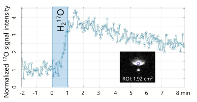
of brain water dynamics in the occipital lobe using 17O MRI:
short- and long-term observations
M. Tomiyasu, H. Sano, Y. Bito, T. Oono, H. Kameda, R.
Kishimoto, T. Omatsu, K. Kudo, T. Obata
National Institutes for Quantum Science and Technology, Chiba, Japan
Impact: This study demonstrates the potential of 17O-MRI
to monitor brain water dynamics in
vivo, capturing both rapid blood flow changes and
longer-term equilibration with interstitial fluids. Findings
support 17O-MRI
as a valuable tool for understanding brain water
distribution over time.
|
|
 |
Computer Number: 51
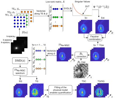
3074. An
efficient post-processing pipeline for improved phase-cycled
23Na Multi-Quantum Coherences MRI
C. Licht, E. Ilicak, F. Boada, V. Jost, M. Guye, F. G.
Zoellner, L. R. Schad, S. Rapacchi
Heidelberg University, Mannheim, Germany
Impact: This pipeline’s algorithms provide the sodium
MRI community with powerful tools for signal separation and
denoising, enabling clearer, more reliable SQ and TQ images
even in challenging conditions, expanding applicability to
clinical and experimental settings.
|
|
 |
Computer Number: 52
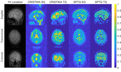
3075. In-vivo
sodium triple quantum (TQ) MR signal extraction using a
single-pulse sequence with single quantum time efficiency at 3T
V. Jost, C. Licht, S. Reichert, D. Zehender, F. Zöllner
Computer Assisted Clinical Medicine, Medical Faculty Mannheim, Heidelberg University, Mannheim, Germany
Impact: The method is readily applicable to any sodium
studies that leverage a multi-echo sodium sequence and
offers therefore, the potential to investigate multi-quantum
coherences, potentially providing richer tissue
characterization than tissue sodium concentration (TSC)
alone.
|
|
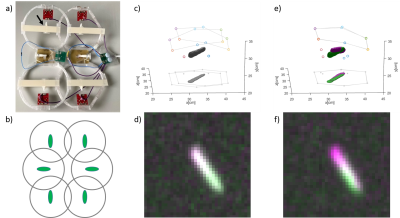 |
Computer Number: 53
3076. Towards
in vivo quantitative 19F MRI in the Spleen
K. Tadjalli Mehr, F. Spreter, S. Reiss, J. Fischer, D. Boll,
A. Özen, C. von zur Mühlen, A. Maier, M. Bock
University Medical Center Freiburg, Freiburg, Germany
Impact: The presented method allows for quantification
of 19F signal decay in non-static large animal organs. While
it already works well on its own, it allows for future
combination with a sensitivity map in the future.
|
|
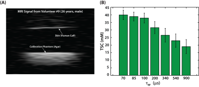 |
Computer Number: 54
3077. Improved
quantification of skin sodium through rapid biexponential
relaxation measurement and signal loss compensation
G. Singh, C. Beaulieu, R. Stobbe
University of Alberta, Edmonton, Canada
Impact: Previous
MRI studies of skin vastly underestimate sodium
concentration. Rapid
biexponential T2 relaxation
measurement enables signal loss compensation to correct skin
sodium concentration values.
|
|
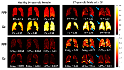 |
Computer Number: 55
3078. Comparison
Of Hyperpolarized 129Xe and 19F Perfluoropropane Multiple-Breath
Washout MRI In Healthy and CF Pediatric Populations
F. Alam, B. Zanette, F. Ratjen, G. Santyr
Hospital for Sick Children, Toronto, Canada
Impact: This work demonstrates how choice of contrast
gas can influence MBW MRI. Understanding differences between
MBW PFP- and Xe-MRI is important as PFP gains interest due
to lower costs and improved clinical translatability for
monitoring treatment progress compared to xenon.
|
|
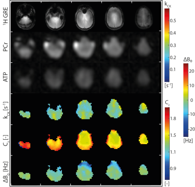 |
Computer Number: 56
3079. Functional
Creatine Kinase Imaging (fCKI) for brain functional and
metabolic imaging
M. Widmaier, A. Kaiser, D. Wenz, Y. Xiao, S-I Lim, Y. Jiang,
L. Xin
EPFL, Lausanne, Switzerland
Impact: A novel functional modality, functional Creatine
Kinase Imaging (fCKI) is introduced. fCKI reveals increased
CK enzyme activity in the occipital lobe and, for the first
time, a 3D activation-map, with activation clusters
predominantly found in the visual cortex.
|
|
 |
Computer Number: 57
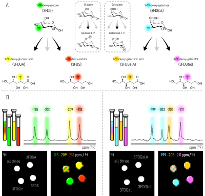
3080. In
vivo Fluorine Metabolic Imaging (FMI) with 3-Fluorodeoxy-Sugars:
A Paradigm Shift in Metabolic Imaging
D. Cohen, B. Subramani, T. Harris, H. Allouche-Arnon, A.
Bar-Shir
Weizmann Institute of Science, Rehovot, Israel
Impact:
FMI with 3FDGal constitutes a robust platform with high metabolite yield, offering an MRI-based approach to studying sugar reduction versus oxidation for the first time. The outlined FMI principles could be extended for imaging additional pathways with other 19F-labeled compounds. |
|
|
Computer Number:
3081. WITHDRAWN |
||
 |
Computer Number: 58
3082. Muscle
Glucose Uptake using Deuterium Metablic Imaging (DMI) at 7T
J. Kaggie, B. Buchignani, P. Ambrosi, P. Cechi, G.
Aringhieri, C. Laustsen, R. Schulte, M. Tosetti
University of Cambridge, Cambridge, United Kingdom
Impact: Deuterated metabolic imaging (DMI) has the
potential to revolutionize the assessment of muscle glucose
metabolism, enabling clinicians to better diagnose and
manage musculoskeletal disorders. This research establishes
baseline methods before applying DMI in musculoskeletal
disorders.
|
|
 |
Computer Number: 59
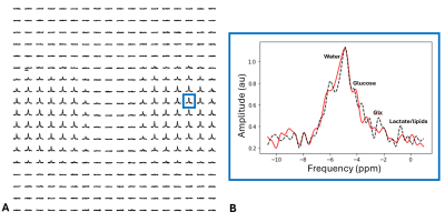
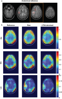
3083. A
Manifold Learning-based Approach for Denoising in Deuterium
Metabolic Imaging
D. Chi, P. Han, H. De Feyter, R. de Graaf, C. Ma
Yale School of Medicine, New Haven, United States
Impact: The LTSA model reduces the noise in DMI signal
and thus improves the estimation of metabolite
concentration. This improvement prospectively allows DMI
with high spatial-temporal resolution, which can assist
tumor diagnosis and treatment response assessment in
clinical settings.
|
|
 |
Computer Number: 60
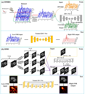
3084. High-Resolution
Deuterium Metabolic Spectroscopy and Imaging with
Self-Supervised Deep Denoising
G. Chen, X. Liu, J. Zhao, S. Wang, P. Sun, L. Frydman, M.
Neeman, X. Zhou, M. Liu, C. Liu, Q. Bao
Innovation Academy for Precision Measurement Science and Technology, Chinese Academy of Sciences, Wuhan, China
Impact: The constructed self-supervised deep denoising
method significantly enhances SNR, enabling high
spatiotemporal resolution DMRS/DMI.
|
|
 |
Computer Number: 61
3085. Measuring
the rapid uptake of 2H in the brain following ingestion of heavy
water
D. Cocking, R. Damion, M. Brook, D. Auer, R. Bowtell
University of Nottingham, Nottingham, United Kingdom
Impact: This study demonstrates the feasibility of using
²H MRI to non-invasively monitor deuterium rapid changes in
the distribution of deuterium label in brain tissues. This
can support research on lipid synthesis, protein turnover,
and other physiological processes with minimal
invasiveness.
|
|
 |
Computer Number: 62
3086. Deuterium
magnetic resonance imaging of tumors after in vivo deuterated
water labeling to low total body water levels.
M. He, J. Duzen, H. Merkle, J. Spernyak, T. Larus, D.
Farthing, N. Buxbaum
Roswell Park Comprehensive Cancer Center, Buffalo, United States
Impact: Systemic administration of deuterated water can
preferentially label tumors for dMRI. This approach has been
tested in animal models, while clinical implementation in
oncology warrants lower doses and shorter labeling
durations. Thus, we tested clinically relevant deuterated
water labeling schemas.
|
|
 |
Computer Number: 63
3087. Design
of a dynamic Sodium (23Na) phantom to evaluate NORDIC on changes
in tissue sodium concentration.
B. Prestwich, S. Francis
University of Nottingham, Nottingham, United Kingdom
Impact: A dynamic phantom to validate the sensitivity to
assessing a spatially defined dynamic change in 23Na
concentration, to mimic changes which may occur in muscle or
brain functional sodium and apply NORDIC denoising to the
data.
|
 Back to Meeting Home
 Back to the Program-at-a-Glance
The International Society for Magnetic Resonance in Medicine is accredited by the Accreditation Council for Continuing Medical Education to provide continuing medical education for physicians.
 View
Presentation Video
View
Presentation Video