Digital Poster
CEST, APT, & NOE
ISMRM & ISMRT Annual Meeting & Exhibition • 10-15 May 2025 • Honolulu, Hawai'i

 |
Computer Number: 33
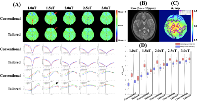
2293. B1
inhomogeneity mitigated chemical exchange saturation transfer
imaging using tailored saturation pulses

J. Tang, Y. Zhang
Key Laboratory for Biomedical Engineering of Ministry of Education, Department of Biomedical Engineering, College of Biomedical Engineering & Instrument Science, Zhejiang University, Hangzhou, China
Impact: A new method to mitigate the influence of B1 inhomogeneity
for CEST imaging is provided. The method has the advantage
of being easy to implement and not increasing the
acquisition duration.
|
|
 |
Computer Number: 34
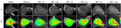
2294. Experimental
study on evaluating renal redox metabolism in renal
ischemia-reperfusion injury using GluCEST imaging with 3.0 T MRI
L. Liu, W. Mi, X. Yu, W. Xing, L. Pan
Third Affiliated Hospital of Soochow University, ChangZhou, China
Impact:
This study demonstrates that GluCEST is a noninvasive and reliable imaging technique that evaluates the renal redox metabolism status after IRI and aids in the early diagnosis and clinical management of renal IRI. |
|
 |
Computer Number: 35
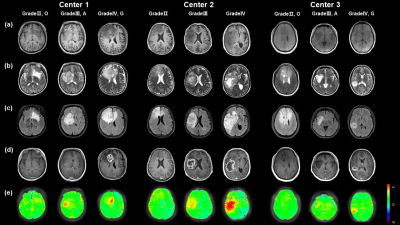
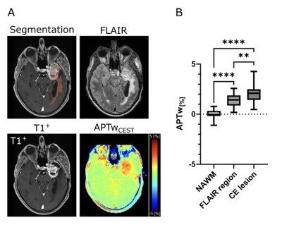
2295. Multicenter
Validation of Amide Proton Transfer Imaging for Molecular and
Grade Classification of Adult-Type Diffuse Gliomas
T. Jiang, M. Wu, J. Hu, Y. Hsu, H. Guo, X. Ma, Y. Liu, Y.
Zhang
Zhejiang University, Hangzhou, China
Impact: This study demonstrates that APT imaging
reliably differentiates adult-type gliomas across centers,
enabling more precise, non-invasive tumor characterization.
It promotes earlier, molecularly informed diagnoses, guiding
personalized treatment strategies and fostering research on
APT imaging's prognostic and therapeutic potential.
|
|
 |
Computer Number: 36
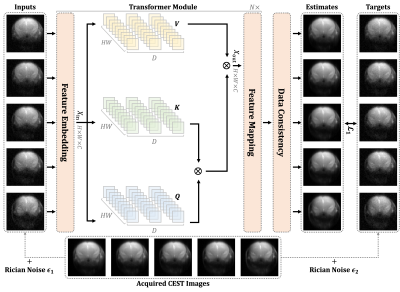
2296. Enhancing
SNR of CEST MRI Through Noise-to-Noise Deep Learning with
K-space Data Consistency
H. LIU, Z. XIA, L. H. LAW, Z. CHEN, J. HUANG, D. SHEN, K.
CHAN
City University of Hong Kong, Hong Kong, China
Impact: Our method can considerably increase SNR of CEST
images without sacrificing image fidelity. After denoising,
the derived CEST maps could more reliably represent
molecular changes in brain regions.
|
|
 |
Computer Number: 37
2297. Mitigation
of T1 impact for unbiased tumor APT MRI using quasi-steady-state
based apparent exchange-dependent relaxation analysis
Z. Liu, Q. Yang, H. Liu, H. Luo, Y. Zheng, W. Cui, D. Luo,
Y. Wu
National Cancer Center/National Clinical Research Center for Cancer/Cancer Hospital & Shenzhen Hospital, Chinese Academy of Medical Sciences and Peking Union Medical College, Shenzhen, China
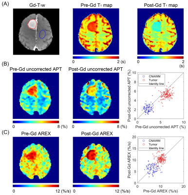
Impact: The QUASS-based AREX analysis can effectively
correct the T1 confoundment
in CEST measurements, facilitating unbiased tumor CEST MRI
at 3 T.
|
|
 |
Computer Number: 38
2298. CEST
contrasts remain stable in normal-appearing brain tissues but
reveal tumor-specific alterations after radiotherapy in glioma
patients
S. Regnery, N. von Knebel Döberitz, F. Kroh, P. Boyd, P.
Menshchikov, S. Graß, C. Bauspieß, M. Ladd, J. Debus, H-P
Schlemmer, A. Korzowski, L. König, D. Paech
Heidelberg University Hospital, Heidelberg, Germany
Impact: CEST contrasts remain unchanged in
normal-appearing brain tissue after radiotherapy but change
inside gliomas. Based on these findings, CEST contrast
changes in gliomas after radiotherapy should be considered
to be tumor-specific, and may be used to support tumor
response assessment.
|
|
 |
Computer Number: 39
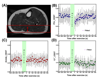
2299. Oxidative
Phosphorylation in Skeletal Muscle Measured by
High-Temporal-Resolution CEST MRI at 3T
L. Ju, T. Samuel, M. Schär, K. Wang, Y. Wu, R. Weiss, J. Xu
Johns Hopkins University, Baltimore, United States
Impact: With temporal resolution comparable to the 31P
MRS technique, UFZ MRI shows great potential as a robust
tool for non-invasive, localized assessment of mitochondrial
function in skeletal muscles, which will assist the
diagnosis of related diseases.
|
|
 |
Computer Number: 40
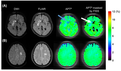
2300. CEST
fluid artifact identification by steady-state signal fitting
(FISS)
X. Yong, L. Zhou, C. Zhang, Y-C Hsu, C. Su, S. Lu, Y. Zhang
Zhejiang University, Hangzhou, China
Impact: The proposed method shows the ability to
identify the fluid compartments on CEST images,which can
help mitigate fluid artifacts in quantitative analysis and
improve image reading.
|
|
 |
Computer Number: 41
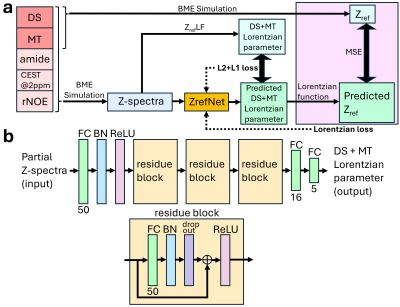
2301. CEST
analysis using a deep residual learning method based on
Bloch-McConnell synthetic training data
S. Zeng, H. Zhang, Z. Wang, J. Wang, P. Cai, J. Huang
the University of Hong Kong, Hong Kong, China
Impact: We
proposed deep
residual networks to predict the Z-spectrum reference signal
(ZrefNet) for CEST quantification. ZrefNet generated
accurate CEST contrasts on simulation data and demonstrated
its feasibility for analyzing in vivo human CEST data of
multiple sclerosis and healthy controls.
|
|
 |
Computer Number: 42
2302. The
Value of Amide Proton Transfer MRI in the Grading of Clear Cell
Renal Cell Carcinoma : Comparison With Diffusion-Weighted
Imaging
X. Yang, H. Gao, C. Shen, Y. Fan, C. Zhang, D. Liu, P. Wu,
L. Han, Y. Bai
Liaocheng People's Hospital, Liaocheng, China
Impact: APTw MRI holds potential as a valuable
supplementary sequence to conventional MRI for the grading
of ccRCC.
|
|
 |
Computer Number: 43
2303. Magnetic
Resonance Fingerprinting for Glutamate Quantification: Towards
the Intermediate Exchange Regime
D. Korenchan, N. Vladimirov, O. Perlman, C. Farrar
Athinoula A. Martinos Center for Biomedical Imaging, Charlestown, United States
Impact: We demonstrate the initial development of
quantitative glutamate imaging using chemical exchange and
fingerprinting, which offers higher spatial resolution than
spectroscopy and can track disease-dependent glutamate
changes in the brain, such as for cancer or
neurodegeneration.
|
|
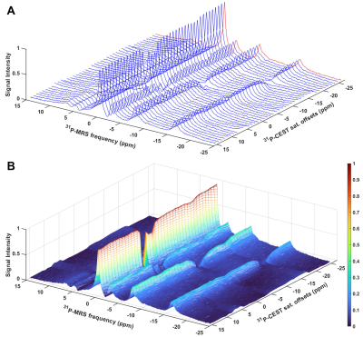 |
Computer Number: 44
2304. 31P-CEST
“matrix” for in vivo rat brain: novel apparent Z spectra
analysis towards sensitive imaging
N. Gao, Y. Wang, J-H Gao, X. Song
Center for Biomedical Imaging Research, Tsinghua University, Beijing, China
Impact: This study demonstrates that 31P-CEST
technique enables the detection of individual metabolites
(several mM) by indirectly detecting the bulk 31P
signal (~20 mM), indicating the potential of 31P-CEST-MRI
for accelerating acquisition and enhancing sensitivity by
removing the spectral dimension.
|
|
 |
Computer Number: 45
2305. Histogram
Analysis of 3D Amide Proton Transfer-weighted MRI for
Distinguishing Brain Abscesses from Cystic High-grade Gliomas
X. Zou, Y. Xu, Z. Wen
Zhujiang Hospital of Southern Medical University, Guangzhou, China
Impact: This study is the first to utilize APTw imaging
to differentiate between brain abscesses and cystic
high-grade gliomas. The results highlight the potential of
APTw as a valuable imaging biomarker, supporting its utility
in improving diagnostic accuracy.
|
|
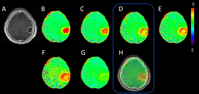 |
Computer Number: 46
2306. APTw-MRI
with additional suppression of fluid compartments (necrosis) and
noise reduction using rescaled MT-correcting CEST metrics
J. Keupp, O. Togao
Philips Innovative Technologies, Hamburg, Germany
Impact: The rescaled metric may provide a standard for
displaying APTw contrast in tumor diagnosis and response
assessment, combining MT-corrected and low noise
quantitative assessment of chemical exchange in solid tumor
areas with extra fluid suppression for efficient clinical
reading.
|
|
 |
Computer Number: 47
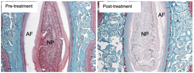
2307. R1rho
Dispersion Imaging to Detect Proteoglycan Loss in the
Intervertebral Discs in Swine Models
M. Christiansen, S. Perez, M. Preul, J. Turner, J. Uribe, E.
Mufson, D. Yang, W. Yoo, J. Gore, R. Dortch, P. Wang
Barrow Neurological Institute, Phoenix, United States
Impact: The study suggested that R1rho dispersion can
detect the proteoglycan loss in spine discs, potentially
making it a promising tool for the early detection of disc
degeneration.
|
|
 |
Computer Number: 48
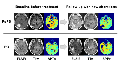
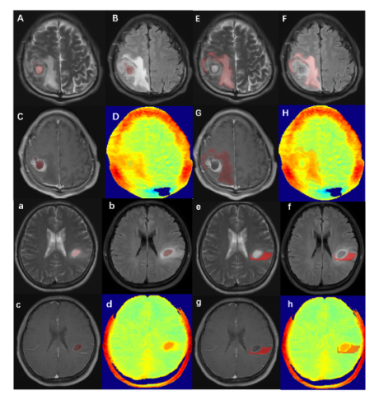
2308. Differentiating
glioma recurrence and pseudoprogression by APTw CEST MRI
K. Karimian-Jazi, N. Enbergs, E. Golubtsov, K. Schregel, J.
Ungermann, H. Fels-Palesandro, D. Schwarz, V. Sturm, J.
Kernbach, D. Batra, F. Ippen, I. Pflüger, N. von Knebel
Doeberitz, S. Heiland, M. Platten, F. Winkler, W. Wick, D.
Paech, M. Bendszus, M. Breckwoldt
University Hospital Heidelberg, Heidelberg, Germany
Impact: This study highlights APTw-CEST MRI's value as
an imaging biomarker, aiding clinicians in differentiating
progression from pseudoprogression. It enables targeted
treatment strategies and opens new research into metabolic
imaging for personalized glioma management, enhancing
patient care and guiding therapy decisions.
|
 Back to Meeting Home
 Back to the Program-at-a-Glance
The International Society for Magnetic Resonance in Medicine is accredited by the Accreditation Council for Continuing Medical Education to provide continuing medical education for physicians.
 View
Presentation Video
View
Presentation Video