Oral
Data Post-Processing
ISMRM & ISMRT Annual Meeting & Exhibition • 10-15 May 2025 • Honolulu, Hawai'i

| 15:45 |
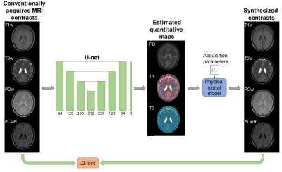 |
1045. Retrospective
relaxometry from conventional contrasts by physics-informed deep
learning: A pilot on Tumor, MS, Stroke and Epilepsy patients
J. van Lune, S. Mandija, M. Schilder, L. Jacobs, J.
Kleinloog, M. Maspero, S. Jacobs, C. van den Berg, A.
Sbrizzi
University Medical Center Utrecht, Utrecht, Netherlands
Impact:
This study demonstrates the use of self-supervised, physics-informed deep learning to generate full-brain quantitative T1- and T2-maps from conventional MRI on a heterogeneous dataset of neurological patients, potentially enabling future applications on large-scale datasets to improve diagnostic tools. |
| 15:57 |
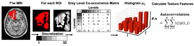 |
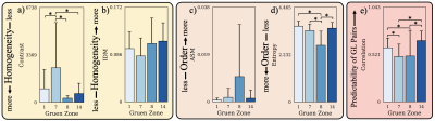
1046. Texture
Features as Sensitive Markers for Early Detection and
Differentiation of Disease Stages in Behavioral Variant
Frontotemporal dementia
B. Akbarian, K. Hett, T. Phan, R. Darby
Vanderbilt University, Nashville, United States
Impact: This study advances the understanding of
frontotemporal dementia by demonstrating the utility of
texture-based MRI features as sensitive biomarkers for early
detection and differentiating between disease stages,
potentially improving diagnosis accuracy and developing
personalized treatment strategies.
|
| 16:09 |
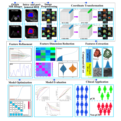 |
1047. Novel
MRI-based Hyper-Fused Radiomics for Predicting Pathologic
Complete Response to Neoadjuvant Therapy in Breast Cancer
Q. Cui, L. Zhou, X. Wang, H. Guan, Z. Kuai
Harbin Medical University Cancer Hospital, Harbin, China
Impact: This research highlights hyper-fused radiomics
as a promising tool in precision oncology, potentially
replacing contrast-based imaging for patients with
contraindications and advancing predictive accuracy in
therapy response.
|
| 16:21 |
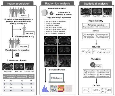 |
1048. Robustness
of MRI radiomics features in abdomen: impact of deep learning
reconstruction and accelerated acquisition
J. Zhong, Y. Xing, Y. Hu, X. Liu, D. Ding, S. Dai, J. Lu, Y.
Song, M. Lu, H. Zhang, W. Yao
Tongren Hospital, Shanghai Jiao Tong University School of Medicine, Shanghai, China
Impact: Deep learning reconstruction and accelerated
acquisition significantly impacts on radiomic features,
necessitating caution to the generalizability when
performing radiomic analysis using images from different
reconstruction algorithms and acquisition protocols.
|
| 16:33 |
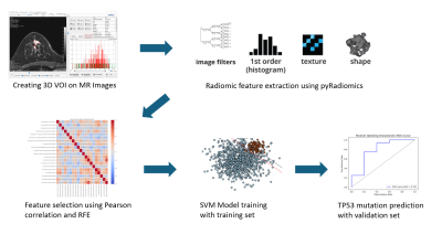 |
1049. Multimodality
MRI radiomics analysis for predicting TP53 mutations in triple
negative breast cancer
J. Hwang, Y. J. Lee, E. Kim, S. H. Kim
Hanyang University, Seoul, Korea, Republic of
Impact: This machine learning-based MRI radiomics model,
trained on multi-center, multi-vendor data, demonstrated
strong predictive performance, enhancing reliability,
generalizability, and patient convenience. It reduces costs
compared to invasive methods and offers broad clinical
applicability across diverse fields.
|
| 16:45 |
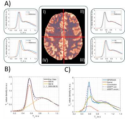 |
1050. Bias
field correction for T1 mapping using phase-cycled bSSFP
N. Plähn, Y. Safarkhanlo, E. Pepper, B. Açikgöz, A.
Mackowiak, G. Bonanno, R. Heule, J. Bastiaansen
Interventional and Pediatric Radiology (DIPR), Inselspital, Bern University Hospital, University of Bern, Bern, Switzerland
Impact: Inaccuracies in T1 quantification from multiple
bias sources, such as transmit field inhomogeneities or
magnetization transfer may be removed simultaneously by
utilizing the known T1 distribution of brain tissues. This
ultimately may enable more objective tissue
characterization.
|
| 16:57 |
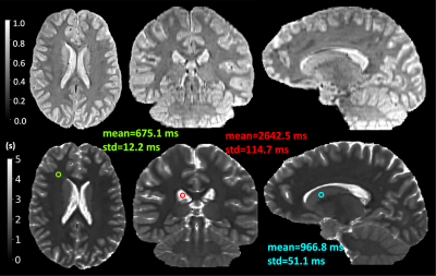 |
1051. High-Resolution
Quantitative T1 Map Estimation for Brain MRI with Clinically
Practical Scan Durations
Z. Huang, H. Ouaalam, O. Afacan, S. K. Warfield, Y. Sui
National Institute of Health Data Science, Peking University, Beijing, China
Impact: We developed a methodology for estimating
high-resolution T1 maps
from a minimal set of low-resolution images acquired in
around 6 minutes, allowing for accurately measuring
quantitative T1 relaxation
times at 0.7mm high isotropic resolution.
|
| 17:09 |
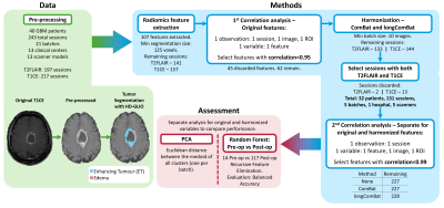 |
1052. Does
Harmonization Impact Radiomics Features in Longitudinal MRI of
Glioblastoma? A Comparison between ComBat and longComBat
M. P. Loureiro, C. Passarinho, A. Matoso, R. Reis Nunes, J.
Maria Moreira, P. Vilela, P. Figueiredo, R. G. Nunes
Instituto Superior Técnico, Universidade de Lisboa, Lisbon, Portugal
Impact: Technical variations greatly influence
MRI-derived radiomics features. We harmonized radiomics
features from longitudinal MRI of Glioblastoma using both
ComBat and longComBat. The latter improved Machine Learning
classification into pre- and post-operative scans.
|
| 17:21 |
 |
1053. MRI-Based
Radiomic Analysis of Trabecular Bone Surrounding Total Hip
Arthroplasty in Femoral Loosening and Asymptomatic Patients
J. Consolini, E. Koretsky, M. Koff, H. Potter
Hospital for Special Surgery, New York, United States
Impact: Image texture analysis may aid in identifying
trabecular bone health, which can indicate an individual’s
risk of aseptic loosening and potential requirement for
revision surgery.
|
| 17:33 |
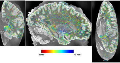 |
1054. A
method for automatic 3D vasculature segmentation in ex vivo MRI
using synthetic data
C. Mauri, E. Chollet, A. Willis, A. Jama, A. Mahmood, A.
Ream, I. Garcia, M. Benlahcen, S. Wood, S. Lin, P. Onta,
N. Tran, X. Zeng, C. Magnain, R. Herisse, E. Garcia
Pallares, M. Hoffmann*, B. Fischl*, Y. Balbastre*
Athinoula A. Martinos Center for Biomedical Imaging, Department of Radiology, Massachusetts General Hospital, Charlestown, United States
Impact: Our method for 3D vessel segmentation in ex
vivo MRI can be used to build a whole-brain vascular
atlas, and study inter-subject variability. It can also be
adapted to microscopy and neuropathology, and to other
tubular structures (axons and fascicles).
|
 Back to Meeting Home
 Back to the Program-at-a-Glance
The International Society for Magnetic Resonance in Medicine is accredited by the Accreditation Council for Continuing Medical Education to provide continuing medical education for physicians.
 View
Presentation Video
View
Presentation Video