Oral
Technical Developments in Musculoskeletal MRI
ISMRM & ISMRT Annual Meeting & Exhibition • 10-15 May 2025 • Honolulu, Hawai'i

Oral
Technical Developments in Musculoskeletal MRI
| 08:15 |
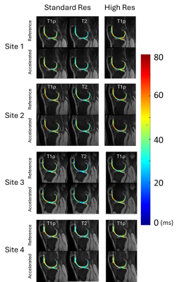 |
1154. Accelerated
T1ρ and T2 High Resolution Imaging: a Multi-vendor Multi-site
study

Z. Zhang, J. Kim, R. Liu, R. Latery, C. Winalski, M.
Yang, J. Liu, T. Link, Q. Peng, M. Samaan, P. Hardy, L.
Ying, X. Li
Cleveland Clinic, Cleveland, United States
Impact: We demonstrated high-resolution fast cartilage
relaxometry imaging is reliable and reproducible, with
excellent agreement between reference and accelerated
imaging, excellent scan-rescan repeatability, and
consistency across sites and vendors. Such techniques will
greatly facilitate the clinical translation of quantitative
knee imaging.
|
| 08:27 |
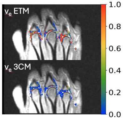 |
1155. DCEMRI
Modelling in the Presence of Ill-perfused Regions
J. Waterton, J. Naish, A. Mahmutovic, L. Edwards, M. Heaton,
M. Tibiletti, L. Nordenmark, G. Parker, J. MacKay
Bioxydyn, MANCHESTER, United Kingdom
Impact: A robust DCEMRI analysis with better face
validity increases confidence in the pathophysiologic
interpretation of any pharmacodynamic changes.
|
| 08:39 |
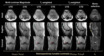 |
1156. Simultaneous
solid and multiple-contrast soft tissue musculoskeletal magnetic
resonance imaging
E. Baccaglini, B-T Vu, N. Kamona, F. Wehrli, C. Rajapakse
University of Pennsylvania, Philadelphia, United States
Impact: DREAMER performs simultaneous imaging of
musculoskeletal solid and soft tissues, potentially
obviating the need for additional CT. It can reduce the use
of ionizing radiation in clinical imaging and remove
logistical complexities related to scheduling examinations.
|
| 08:51 |
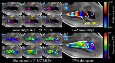 |
1157. A
new technique for detecting waves parallel to muscle fibers in
MR elastography: Application to the supraspinatus muscle
D. Ito, T. Numano, T. Habe, T. Nozaki, Y. Ishihara, J.
Tsuzaki, M. Hase, M. Arai, M. Jinzaki
Keio University Hospital, Tokyo, Japan
Impact: Our technique enhances the reliability of
skeletal muscle MRE by extracting waves along the muscle
fiber direction and automating stiffness measurements. This
advancement enables accurate assessment of pathological and
physiological changes, supporting early diagnosis and better
assessment of musculoskeletal disorders.
|
| 09:03 |
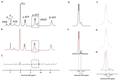 |
1158. NAD
in skeletal muscle at 3 T: impact of age and disease
H. Reyngoudt, B. Matot, V. Henriet, Y. Fromes, B. Marty
Institute of Myology, Paris, France
Impact: Since NAD is abundant in living cells and
sensitive to change (age, disease, exercise,
supplementation) and 31P
MRS allows its quantification, this metabolite can serve as
a potential quantitative MR biomarker in skeletal muscle
studies.
|
| 09:15 |
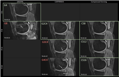 |
1159. Cartesian
vs. non-cartesian under-sampling for high resolution 3D double
echo steady state (DESS) knee imaging at 7 T
T. Marth, A. Marth, G. Kajdi, D. Paul, R. Sutter, D. Nanz,
C. von Deuster
Balgrist Campus AG, Zurich, Switzerland
Impact: The results suggest significant advantages of
non-cartesian versus cartesian undersampling strategies in
DESS imaging with high acceleration factors at 7T. The short
acquisition times may allow clinical high-resolution 3D knee
cartilage imaging, e.g., for quantification of cartilage
morphology.
|
| 09:27 |
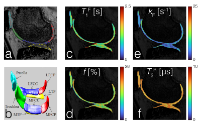 |
1160. Advanced
In-Vivo 3D Quantitative Magnetization Transfer Imaging of
Cartilage Extracellular Matrix of the Whole Knee Joint
A. Jang, Z. Stewart, M. Jarraya, F. Liu
Athinoula A. Martinos Center for Biomedical Imaging, Charlestown, United States
Impact:
BTS showed spatial variation and regional features of qMT parameters in a healthy model, presenting great potential in providing 3D relaxometry and qMT imaging biomarkers to characterize whole knee cartilage extracellular matrix, which requires further investigation using groupwise analysis. |
| 09:39 |
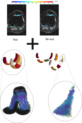 |
1161. Cartilage
shape-informed 3D gray level co-occurrence matrix analysis of
DESS T2 maps
V. Janacova, P. Szomolanyi, D. Sitarcikova, S. Trattnig, V.
Juras
Medical University of Vienna, Vienna, Austria
Impact: This study demonstrates the high repeatability
of cartilage shape informed 3D GLCM features. Since
cartilage structure varies in directions perpendicular and
parallel to the bone-cartilage interface, accurately
assessing texture in these anatomically relevant
orientations is clinically important.
|
| 09:51 |
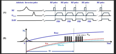 |
1162. Fast
Three-Dimensional Short TR Adiabatic Inversion Recovery
Ultrashort Echo Time (STAIR-UTE) Imaging of Cortical Bone
M. Daskareh, M. Carl, A. Suprana, J. Chen, J. Lo, S. Jerban,
E. Chang, C. Chung, Y. Ma, J. Du
UC San Diego, San Diego, United States
Impact: STAIR-UTE allows fast direct imaging of cortical
bone at various anatomical sites with high positive
contrast, with potential for routine clinical applications
|
| 10:03 |
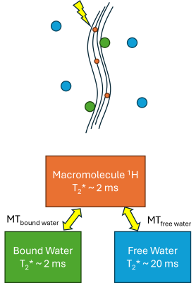 |
1163. Optimization
and Mechanisms of UTE-Magnetization Transfer (MT) contrast in
the ACL
E. Argentieri, M. Han, S. Majumdar, P. Larson
University of California - San Francisco, San Francisco, United States
Impact: These parameters and understanding of MT in the
ACL using UTE MRI will be beneficial to better interpret MT
measurements as well as for designing improved imaging
protocols that will be more sensitive to degeneration.
|
 Back to Meeting Home
 Back to the Program-at-a-Glance
The International Society for Magnetic Resonance in Medicine is accredited by the Accreditation Council for Continuing Medical Education to provide continuing medical education for physicians.
 View
Presentation Video
View
Presentation Video