Oral
Advances in Brain Tumor Imaging: From Diagnosis to Therapy Monitoring
ISMRM & ISMRT Annual Meeting & Exhibition • 10-15 May 2025 • Honolulu, Hawai'i

Oral
Advances in Brain Tumor Imaging: From Diagnosis to Therapy Monitoring
| 13:30 |
Introduction
Sonoko Oshima
|
|
| 13:42 |
 |
0880. Perfusion
and Time of Exchange Measurements in IDH Mutational Subgroups of
Gliomas Using BBB-ASL

G. Turhan, A. Çetin, B. Esteves Padrela, A. Mahroo, S.
Konstandin, D. Hoinkiss, N. Breutigam, V. Keil, A. Ersen
Danyeli, K. Özduman, K. Eickel, H. Mutsaerts, M.
Günther, J. Petr, A. Dinçer, E. Ozturk-Isik
Bogazici University, İstanbul, Turkey
Impact: Our findings underscore the promising potential
of multi-echo BBB-ASL in evaluating BBB integrity and
identifying IDH mutational subgroups of gliomas.
|
| 13:54 |
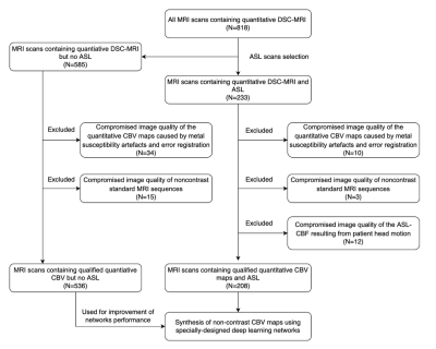 |
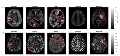
0881. Non-contrast
Cerebral Blood Volume Images Synthesis from Arterial Spin
Labeling and Non-contrast Standard MRI Using Deep Learning
Network
B. Wang, Y. Pan, J. Xin, C. Wang, J. He
Qilu Hospital of Shandong University, Jinan, China
Impact: Patients undergoing radiochemotherapy with
fragile vessels or adverse reactions to gadolinium contrast
could benefit from synthetic non-contrast CBV methods.
|
| 14:06 |
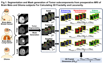 |
0882. Geometry
Matters: Quantifying 3D-Fractality and Lacunarity in Tumor
Subcomponents Distinguishes Between Brain Metastases and Gliomas
N. Yadav, J. Baibhav, V. Tiwari
Indian Institute of Science Education and Research Berhampur, Berhampur, India
Impact: This study introduces fractal-based
tumor-geometry-metrics as innovative, non-invasive
imaging-signature distinguishing brain-metastases
(arising-from-breast and -lung cancers) and gliomas.
Integrating 3D-fractality and lacunarity measurements of
tumor-subcomponents in machine-learning-models yielded
high-accuracy, precise-differentiation between
brain-metastases and gliomas, thus reducing
biopsy-dependency, enhancing
noninvasive-differential-diagnosis, and prognostication.
|
| 14:18 |
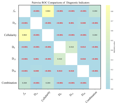 |
0883. Feasibility
of Time-Dependent Diffusion MRI-Based Indicators for Identifying
MGMT Promoter Methylation in High-Grade Gliomas
Q. Wang, Y. Xie, Y. Zhang, X. Liang, Y. Fu, C. Zhang, F.
Thorsten, M. Chen
The 6th Medical Center, Chinese PLA General Hospital, Beijing, China, Beijing, China
Impact: Our findings may help enabling non-invasive
diagnostics for patients with HGGs by providing reliable
prognostic tools and may inspire further research into
MRI-based techniques for characterizing tumors as well as
help guide clinical decision-making for patients unsuitable
for biopsy.
|
| 14:30 |
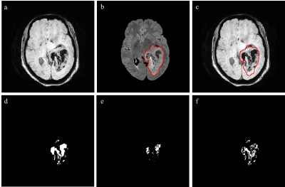 |
0884. Differentiating
hemorrhage from tumor vasculature in SWI-MRI images using Swin
UNETR: Applications in Glioma Grading
S. Maurya, M. Sheikh, R. Gupta, A. Singh
Indian Institute of Technology Delhi, New Delhi, India
Impact: This study presents a method for automatically
segmenting IV from SWI images using Swin UNETR without
multi-echo SWI or R2* maps, enhancing efficiency in
resource-limited settings, showing high accuracy in
classifying gliomas. It addresses the subjectivity inherent
in manual methods.
|
| 14:42 |
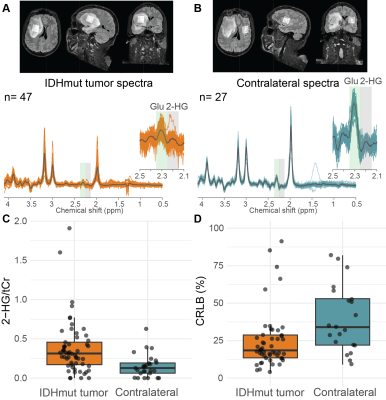 |
0885. Pitfalls
in unedited 2-HG MRS detection with optimized TE at 3T
S. Alcicek, D. Simicic, L. Blair, M. Saint-Germain, H.
J. Zöllner, C. W. Davies-Jenkins, M. Holdhoff, J.
Laterra, C. Bettagowda, K. C. Schreck, D. D. Lin, P. B.
Barker, D. O. Kamson, G. Oeltzschner
The Johns Hopkins University School of Medicine, Baltimore, United States
Impact: This study highlights that false-positive 2-HG
MRS detection can occur in normal-appearing brain tissue and
identifies signal overlap (with GABA, acetone, etc.) as the
primary reason. 2-HG estimates should be interpreted
carefully in clinical context, including dietary preferences
or interventions.
|
| 14:54 |
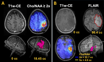 |
0886. Personalized
brain tumor radiation therapy planning pipeline based on
spectroscopic MRI using the Brain Imaging Collaboration Suite
(BrICS)
A. Trivedi, K. Ramesh, A. Giuffrida, S. Sheriff, L.
Cooper, B. Weinberg, S. Ahn, V. Khalilzad Sharghi, A.
Maudsley, J. Alger, B. Soher, H. Shim
Emory University School of Medicine, Atlanta, United States
Impact: PyMIDAS, a Python version of the existing
IDL-based pipeline, accelerates and simplifies spectroscopic
MRI-based brain tumor treatment planning, enabling clinical
workflow integration. Its improved computational efficiency
and flexibility support broader adoption of advanced
spectroscopic MRI for brain tumor imaging.
|
| 15:06 |
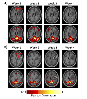 |
0887. First
demonstration of resting-state functional MRI on a 1.5T MR-Linac
to assess functional connectivity in glioblastoma patients
E. Almasri, L. Lawrence, J. Stewart, M. Ruschin, A.
Theriault, J. Detsky, S. Myrehaug, C-L (. Tseng, H.
Soliman, A. Sahgal, A. Lau
University of Toronto , Toronto , Canada
Impact: We showed that frequent resting-state functional
imaging on MRI-linear accelerators is feasible, with high
within-patient and low across-patient session similarity.
These findings suggest a potential for tracking individual
functional network changes during radiotherapy to guide
treatment adaptation in glioblastoma patients.
|
| 15:18 |
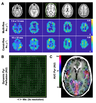 |
0888. Multi-resolution
hyperpolarized 13C EPI with atlas-based prescription for
metabolic imaging of human gliomas
A. Autry, J. Gordon, H-Y Chen, Y. Kim, J. Villanueva-Meyer,
S. Chang, J. Clarke, N. Oberheim-Bush, D. Xu, J. Lupo, P.
Larson, D. Vigneron, Y. Li
University of California San Francisco, San Francisco, United States
Impact: This project improved the assessment of
glycolytic metabolism in patients with gliomas using
multi-resolution EPI that provides a 4-fold increase in
hyperpolarized [1-13C]pyruvate
resolution, while also leveraging automatic atlas-based
prescription for consistent volumetric coverage.
|
 Back to Meeting Home
 Back to the Program-at-a-Glance
The International Society for Magnetic Resonance in Medicine is accredited by the Accreditation Council for Continuing Medical Education to provide continuing medical education for physicians.
 View
Presentation Video
View
Presentation Video