Oral
Advances in White Matter Imaging
ISMRM & ISMRT Annual Meeting & Exhibition • 10-15 May 2025 • Honolulu, Hawai'i

| 15:45 |
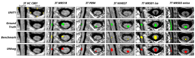 |
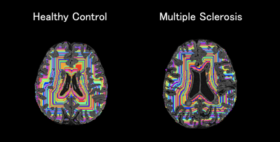
0625. Automatic
Multiple Sclerosis Lesion Segmentation in the Spinal Cord on 3T
and 7T MP2RAGE images
N. Laines Medina, S. Mchinda, B. Testud, S. Demortière, M.
Chen, G. Nair, D. Reich, C. Granziera, C. Tsagkas, V.
Callot, J. Cohen-Adad
Aix Marseille Univ, CNRS, CRMBM, Marseille, France
Impact: This study presents a deep-learning-based method
for MS lesion segmentation in the SC, which enhances
diagnostic accuracy, reduces segmentation time, and offers
lower variability compared to manual approaches,
demonstrating significant potential to impact clinical
practice and improve routine MS diagnosis
|
| 15:57 |
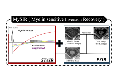 |
0626. Discriminating
White/Gray matter in Cervical Spinal Cord by Myelin-Sensitive
Inversion Recovery with Deep Learning Reconstruction at 1.5T MRI
K. Nitta, H. Yokota, T. Sada, R. Kurosawa, I. Nakanishi, H.
Sato, K. Matsumoto, T. Namiki, M. Yoneyama, T. Iimori, T.
Uno
Chiba University Hospital, Chiba, Japan
Impact:
This research advances the field of myelin-specific MR imaging by demonstrating the feasibility of MySIR at 1.5T MRI with deep learning-based reconstruction. This technique provides an efficient way to visualize myelin, potentially improving the diagnosis of neurological diseases. |
| 16:09 |
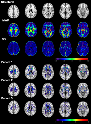 |
0627. Myelin
water imaging in adolescent and young adult cancer patients and
cognitive impairment: A feasibility longitudinal case-control
study
E. J. Lim, C. Y. J. Goh, C. J. Tan, W. Lee, P. H. Chai, M.
F. B. Harunal Rashid, A. Chan, S. Hartono, L. L. Chan
Duke-NUS Medical School, Singapore, Singapore
Impact: Myelin water fraction is a complementary
quantitative marker to fractional anisotropy, detecting
white matter dysmyelination in post-chemotherapy adolescent
and young adult cancer patients.
|
| 16:21 |
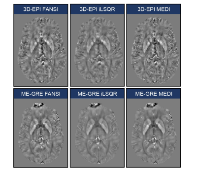 |
0628. A
Comparative Analysis of Different QSM Reconstruction Approaches
for the Characterization of Multiple Sclerosis Pathology

A. Cagol, D. Gkotsoulias, M. Ocampo-Pineda, P-J Lu, X.
Chen, M. Weigel, J. Kuhle, L. Kappos, M. P. Sormani, C.
Granziera
Research Center for Clinical Neuroimmunology and Neuroscience Basel (RC2NB), University Hospital and University of Basel, Basel, Switzerland
Impact: QSM demonstrated substantial agreement across
various acquisition and reconstruction methods and
correlated with MS severity. The different QSM
implementations exhibited small variations in sensitivity to
pathological changes, underscoring the necessity for
standardized protocols to enhance clinical application.
|
| 16:33 |
 |
0629. Periventricular
Gradient of Tissue Microstructure in Patients with MS, NMOSD,
and MOGAD
M. Nakaya, A. Hagiwara, Y. Hoshino, Y. Tomizawa, W. Uchida,
T. Sekine, N. Hara, Y. Tsukamoto, J. Kikuta, S. Kamio, C.
Andica, K. Kamagata, M. Hori, A. Wada, O. Abe, N. Hattori,
S. Aoki
Juntendo University, Tokyo, Japan
Impact: Periventricular gradient of myelin content in MS
is distinct from NMOSD and MOGAD. The correlation between
myelin gradients and clinical measures in MS provides
insights into disease progression and may guide personalized
treatment strategies.
|
| 16:45 |
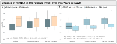 |
0630. Accelerated
mI/tNAA Increase in Normal-Appearing WM of RRMS with
Paramagnetic Rim Lesions: A 7T MR Spectroscopic Imaging 2-year
Follow-Up Study
A. Zöchner, W. Bogner, A. Dal-Bianco, B. Strasser, G.
Hangel, P. Rommer, E. Niess
Medical University of Vienna, Vienna, Austria
Impact: Results revealed faster increase in mI/tNAA in
the NAWM of patients with >1 PRL, which may indicate
elevated inflammation. This could explain the transition
from relapsing-remitting MS to progressive forms and
identify mI/tNAA as potential biomarker for MS progression.
|
| 16:57 |
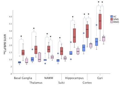 |
0631. Uncovering
fibrin(ogen) deposition in multiple sclerosis brain by in vivo
64Cu-FBP8 MR-PET imaging
C. Treaba, C. Catana, E. Klawiter, S. Huang, R. Bakshi, A.
Misscioscia, E. Barbuti, R. Bomprezzi, J. Sloane, S. Tauhid,
G. Arabasz, P. Caravan, C. Mainero
Athinoula A. Martinos Center for Biomedical Imaging, Massachusetts General Hospital, Boston, United States
Impact: 64Cu-FBP8
MR-PET imaging allows in vivo quantification of fibrin(ogen)
levels and could aid in predicting MS disease outcomes and
improve patient management strategies. Further research is
needed to validate its use as a reliable biomarker in
clinical practice.
|
| 17:09 |
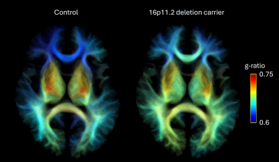 |
0632. Alterations
in white matter tract myelination in carriers of copy number
variations at the 16p11.2 locus

W. D. Lu, C-O Martin, K. Jizi, M. Nelson, S. Jacquemont,
C. Tardif
McGill University, Montreal, Canada
Impact: This quantitative MRI study demonstrates for the
first time that 16p11.2 CNV carriers exhibit altered
myelination of their white matter tracts. Future work will
investigate the association between network myelination and
brain function.
|
| 17:21 |
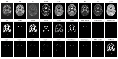 |
0633. Microstructure
fingerprints of white matter hyperintensities in the aging brain
A. Bussy, C. Cathala, F. Kherif, A. Lutti, B. Draganski
Inselspital, Bern, Switzerland
Impact: This study offers unique insights into white
matter hyperintensities (WMH), demonstrating characteristic
patterns of myelin and axonal loss. Our findings highlight
the importance of advanced imaging techniques to better
understand the complexities of WMH and their impact on brain
health.
|
| 17:33 |
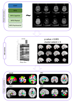 |
0634. Microstructural
and susceptibility alterations in white matter and cerebellum
are linked to fatigue and cognitive dysfunction in long COVID
E. Grosso, N. Kozhanova, M. Gaviraghi, E. Lupi, A.
Monteverdi, M. Yiannakas, A. Ricciardi, M. Battiston, F.
Grussu, R. Samson, F. Prados, B. Kanber, M. Shatila, M.
Zandi, C. Tur, E. D'Angelo, F. Palesi, C. Gandini
Wheeler-Kingshott
University of Pavia, Pavia, Italy
Impact:
This work impacts on our understanding of fatigue and cognitive impairment, impactful symptoms of the long-COVID syndrome, emphasizing the role of diffusion MRI and susceptibility mapping models in detecting microstructural and susceptibility alterations voxel-wise. |
 Back to Meeting Home
 Back to the Program-at-a-Glance
The International Society for Magnetic Resonance in Medicine is accredited by the Accreditation Council for Continuing Medical Education to provide continuing medical education for physicians.
 View
Presentation Video
View
Presentation Video