Power Pitch
Structure-Function Relations in MSK
ISMRM & ISMRT Annual Meeting & Exhibition • 10-15 May 2025 • Honolulu, Hawai'i

Power Pitch
Structure-Function Relations in MSK
| 13:30 |
 |
Screen Number: 26
0962. Patellar
Metabolic and Compositional Markers from 18NaF PET/MRI are
associated with Whole Organ Magnetic Resonance Imaging Scores
and OA-status
R. Bhattacharjee, V. Kreutzinger, Z. Akkaya, K. Ziegeler, E.
Bahroos, M. Han, V. Pedoia, R. Souza, S. Majumdar
University of California, San Francisco (UCSF), San Francisco, United States
Impact:
Presence and extent of morphological bone-marrow-edema, subchondral-cyst, and cartilage-lesion in the patella, and their relationship with peak SUV,T1p, and T2 in patellar subregions in multiple OA subgroups can help elucidate bone remodeling, cartilage degradation and their manifestation in radiographic signs. |
| 13:32 |
 |
Screen Number: 27
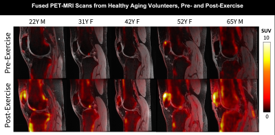
0963. The
Aging Knee: Changes in Bone Metabolic Activity & Cartilage T2
Relaxation Times Measured Using [18F] NaF PET-MRI

A. Goyal, Y. Vainberg, R. Shalit, J. Asay, L. Watkins,
A. Gatti, Y. S. Song, B. Haddock, F. Kogan
Stanford University, Stanford, United States
Impact: This study demonstrates the potential of a
functional [18F]NaF PET-MRI ‘stress test’ for joint health,
offering region-specific joint biomarkers. This work
emphasizes the role of PET-MRI in improving understanding of
how aging-related changes in the joint contribute to
osteoarthritis.
|
| 13:34 |
 |
Screen Number: 28
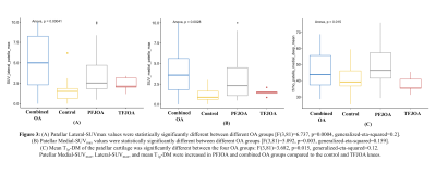
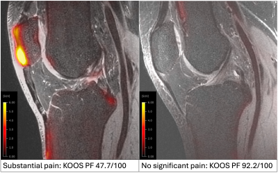
0964. Entheseal
bone remodeling in the anterior patella is associated with worse
functional knee outcomes – new insights from [18F]NaF-PET MRI
K. Ziegeler, V. Kreutzinger, R. Bhattacharjee, E. Hammond,
E. Bahroos, Z. Akkaya, T. Link, R. Souza, S. Majumdar
University of California San Francisco, San Francisco, United States
Impact: Bone remodeling at the enthesis of the patellar
tendon quantified by [18F]-NaF PET-MRI is related to pain
and other patient reported outcomes and may offer inroads to
understanding the poor correlation between structural
lesions and symptoms in OA.
|
| 13:36 |
 |
Screen Number: 29
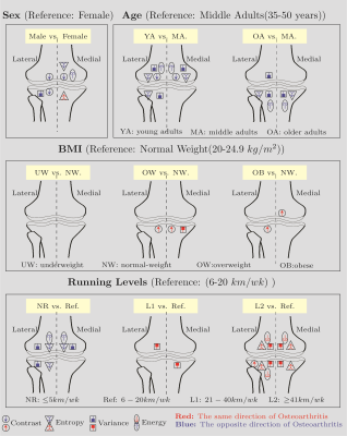
0965. T2
Texture Analysis of Knee Cartilage in Healthy Individuals:
Impact of Regular Running and Demographics
V. Ghodrati, D. Vilimek, J. Uchytil, D. Jandacka, M. T
Nieminen, V. Casula
Research Unit of Health Sciences and Technology, University of Oulu, Oulu, FINLAND, Oulu, Finland
Impact: These results may advance machine learning
models using non-invasive T2 texture analysis to identify
osteoarthritis risk, promoting research into preventive
strategies and enhancing monitoring and early interventions
for joint health through improved understanding of cartilage
changes.
|
| 13:38 |
 |
Screen Number: 30
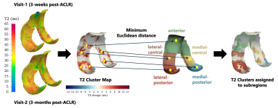
0966. Subregion-Specific
T2-Cluster Analysis Reveals Early Cartilage Changes in Anterior
Cruciate Ligament Reconstructed Knees
V. Barriobero, M. Black, K. Young, J. Asay, S. Sherman,
F. Kogan, G. Gold, B. Hargreaves, A. Chaudhari, A.
Gatti, A. Pai S
Harvard University, Cambridge, United States
Impact: T2-clusters are better at detecting
spatiotemporal variations in T2 within femoral cartilage
subregions compared to bulk mean T2. T2-clusters are
consistent with patterns of full-thickness defects observed
in osteoarthritic cartilage, suggesting its potential
application in monitoring cartilage health post-ACLR.
|
| 13:40 |
 |
Screen Number: 31
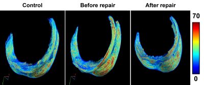
0967. 3D
T2* Mapping of Articular Cartilage at 7 Tesla in Patients Before
and After Medial Meniscus Posterior Root Tear Repair
K. Knutsen, C. Steinberger, E. Hedayati, A. Lamba, S. Zbyn,
L. Tollefson, T. Takahashi, G. Metzger, R. LaPrade, A.
Kajabi, J. Ellermann
University of Minnesota , Minneapolis , United States
Impact: Post-operative monitoring of articular cartilage
with 3D T2* mapping at 7T, which benefits from an enhanced
signal-to-noise ratio, provides valuable insights into
healing progression, guides timely treatments and
potentially helps prevent osteoarthritis.
|
| 13:42 |
 |
Screen Number: 32
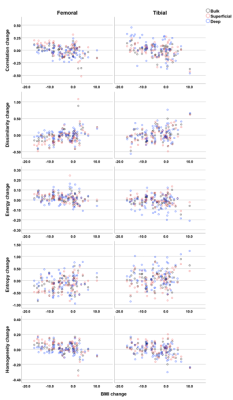
0968. Positive
Effect of Weight Loss on Knee Articular Cartilage – a Three-year
Follow-up Using Texture Analysis of T2 Relaxation Time Maps
E. Mäkelä, V. Casula, A. Kemppainen, M. Haapea, M. Nieminen
University of Oulu, Oulu, Finland
Impact: The results support prior evidence for the
benefits of weight loss for cartilage health and are
important in the context of osteoarthritis prevention and
treatment.
|
| 13:44 |
 |
Screen Number: 33
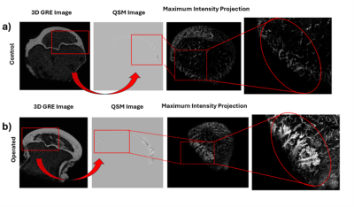
0969. QSM
of epiphyseal cartilage neovascularization following femoral
head ischemia: a retrospective 9.4T piglet model study
A. Shekarabi, E. Buko, S. Parvaze Pathan, C. Johnson
University of Minnesota, Minneapolis, United States
Impact: This work demonstrates improved visualization
and characterization of neovascularization following
ischemic injury and revascularization of epiphyseal
cartilage using the recently described QSM processing
pipeline. This tool may be useful for studying the
pathogenesis of LCPD and related developmental joint
disorders.
|
| 13:46 |
 |
Screen Number: 34
0970. Quantitative
Assessment of Patellar Tendon Recovery and Anterior Cruciate
Ligament Graft Quality using Bi-exponential UTE-T1ρ Mapping
H. Lise de Moura, M. Keerthivasan, P. Su, G.
Koerzdoerfer, M. Alaia, R. Regatte
NYU Langone Health, New York, United States
Impact: The sequence demonstrates potential as a tool
for assessing maturity of ACL grafts. It can help understand
the recovery of PT, the quality of ACL reconstruction, and
risk of post-traumatic osteoarthritis. This knowledge could
improve surgical techniques and rehabilitation protocols.
|
| 13:48 |
 |
Screen Number: 35
0971. Gait
influences changes in T2 clusters in an ACL reconstructed
population 2-3 years post-surgery
J. Asay, A. Pai S, J. He, A. Williams, G. Gold, A. Gatti, B.
Hargreaves, C. Chu
Stanford University, Stanford, United States
Impact: T2-cluster
analysis combined with biomechanics may offer new insights
into early cartilage focal changes in areas of loading that
could otherwise be missed using the standard average
regional T2 relaxation
times, potentially providing target areas for future
therapeutics.
|
| 13:50 |
 |
Screen Number: 36
0972. Quantitative
UTE Imaging of the Meniscus Across Different Zones: A Study on
Knee Osteoarthritis
A. Suprana, H. D. Chae, M. Daskareh, S. H. Shin, J.
Athertya, R. Sah, E. Chang, J. Du, Y. Ma
University of California, San Diego, La Jolla, United States
Impact: This study, to our knowledge is the first to
employ UTE-T1, UTE-MT modeling, UTE-AdiabT1ρ, and UTE-T2*to
investigate compositional changes of each of the 3 vascular
zones of the meniscus due to osteoarthritis.
|
| 13:52 |
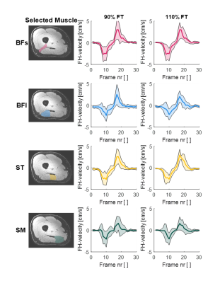 |
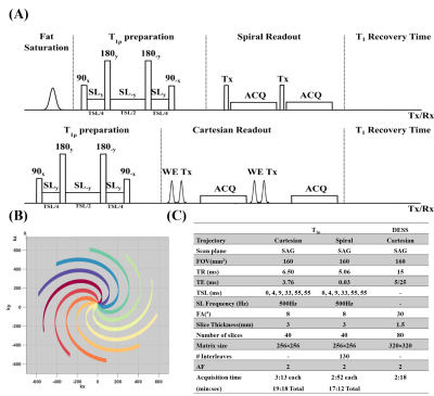
Screen Number: 37
0973. Time-resolved
3D-PC MRI measurements of the upper leg muscles during dynamic
knee flexion: the effect of the fatigue threshold
L. Vos, S. Rauh, H. Kan, A. Nederveen, G. Strijkers, M.
Hooijmans
Amsterdam UMC, Amsterdam, Netherlands
Impact: Exercising slightly above the fatigue threshold
does not impact 3D PC-MRI derived FH velocity data for the
hamstring muscles compared to below-threshold levels,
suggesting that muscle fatigue may have limited effect.
However, further analysis of strain-rate and strain are
required.
|
| 13:54 |
 |
Screen Number: 38
0974. Microstructure
and muscle strength in older adults: a time-dependent diffusion
study
V. Mazzoli, S. Rao, D. Long, S. Kadam, G. Lemberskiy, T.
Feiweier, G. Koerzdoerfer, D. Novikov, E. Fieremans, S.
Coelho
NYU Langone, New York, United States
Impact: The apparent diameter a,
obtained by modeling time-dependent diffusion data combined
with RPBM modeling, is strongly associated to muscle
strength in older adults, and is therefore a promising tool
to study early signs of muscle atrophy and sarcopenia.
|
| 13:56 |
 |
Screen Number: 39
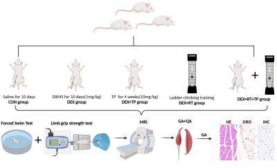
0975. IDEAL-IQ
and BOLD MRI Assessment of Testosterone and Resistance Training
Effects on Fat Infiltration and Blood Perfusion in Sarcopenic
Rats
X. kui, X. Huang, Y. Huang, L. Nie, B. He
The First Affiliated Hospital of Kunming Medical University, Kunming, China, China
Impact: This combined RT and T treatment demonstrates a
potential therapeutic approach for sarcopenia, effectively
enhancing muscle structure and function. IDEAL-IQ and BOLD
MRI offer reliable, noninvasive methods for tracking these
muscle changes in research and clinical settings.
|
| 13:58 |
 |
Screen Number: 40
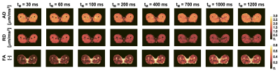
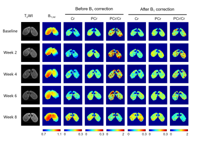
0976. Investigation
of the feasibility of CEST MRI in early detecting diabetic
muscle injuries in a rabbit model at 5 T
Q. Wu, Z. Huang, H. Tang, Y. Wu
Shenzhen Institute of Advanced Technology, Chinese Academy of Sciences, Shenzhen, Guangdong , China
Impact: The significantly increased Cr and reduced
PCr/Cr occurred before structural alterations, suggesting
the feasibility of CEST MRI in early detecting diabetic
muscle damage at the energy metabolism level.
|
| 14:00 |
 |
Screen Number: 41
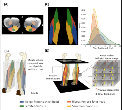
0977. Hamstring
Muscle Architecture and Microstructure Changes Following 9-weeks
of Nordic Hamstring Exercise Training
A. Pai S, M. Andrews, R. Gurchiek, P. Pincheira, M.
Barbieri, T. Friedrich, F. Kogan, G. Gold, G. Lichtwark,
V. Mazzoli, S. Delp, A. Chaudhari
Stanford University, Stanford, United States
Impact: This study examines architectural and
microstructural adaptations of the hamstrings following
9-weeks of Nordic hamstring exercise training. Findings
reveal significant, but non-uniform hypertrophy among
hamstrings accompanied by increase in length and size of the
muscle fibers, advancing injury prevention strategies.
|
| 14:02 |
 |
Screen Number: 42
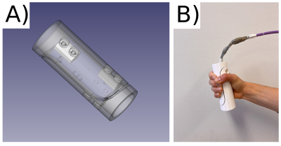
0978. Development
of an open-source, MRI-compatible grip force sensor for dynamic
muscle MRI
S. Räuber, M. Maggioni, P. Panos, F. Santini
University of Basel, Allschwil, Switzerland
Impact: The low-cost, open-source grip force sensor
demonstrated excellent MRI compatibility and therefore
appears to be suitable for use with dynamic muscle MRI
methods to characterise forearm flexors, with potential
applications in the study of myotonic dystrophy.
|
| 14:04 |
 |
Screen Number: 43
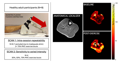
0979. Repeatability
and sensitivity of the skeletal muscle response to in-scanner
isometric exercise with vPIVOT MRI
E. Englund, J. Reusch, A. Barker
University of Colorado, Anschutz Medical Campus, Aurora, United States
Impact: Dynamic measurement of skeletal muscle perfusion
in response to in-scanner exercise with vPIVOT MRI was
repeatable and sensitive to varied exercise intensity in
healthy adults. These results will help to facilitate
comparison in future studies of patient populations.
|
| 14:06 |
 |
Screen Number: 44
0980. Relationship
between function and MRI biomarkers in tongue muscles

E. Vermeulen, P-Y Baudin, Y. Fromes, B. Matot, E.
Giacomini, M. Lapert, J-Y Hogrel, B. Marty
Institute of Myology, Paris, France
Impact: The use of quantitative fat fraction and water
T2 imaging in tongue muscles in addition to functional
testing represents a promising approach for evaluating these
muscles.
|
| 14:08 |
 |
Screen Number: 45
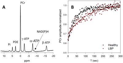
0981. Multifidus
muscle mitochondria function in those with non-specific chronic
low back pain
D. Cawley, S. Adnani, A. Tickal, T. Denney, A. Bashir
Edward Via College of Osteopathic Medicine, Auburn, United States
Impact: We, for the first time, demonstrated the
application of 31P MRS for measurement of mitochondrial
function in lumbar multifidus muscle. Impaired ATP
production in weak/underused multifidus muscle might be
early markers of unspecified lower back pain.
|
| 14:10 |
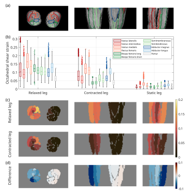 |
Screen Number: 46
0982. 4D
Time-Resolved Strain Tensor Analysis Using Spectro-Dynamic MRI
Reveals Muscle Activation Patterns
M. van Riel, D. Heesterbeek, R. Sheombarsing, M. Froeling,
T. van Leeuwen, C. van den Berg, A. Sbrizzi
UMC Utrecht, Utrecht, Netherlands
Impact: Time-resolved 4D strain analysis using
Spectro-Dynamic MRI enables the measurement of in vivo
biomechanics in a dynamic setting. This creates an
opportunity for new ways to investigate the functioning and
performance of active muscles.
|
| 14:12 |
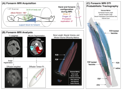 |
Screen Number: 47
0983. Investigating
Forearm Muscle Adaptations in Hemiparetic Cerebral Palsy Using
Diffusion MRI: Structural Correlates of Grip Strength Deficits
D. Joshi, A. Hruby, J. Dewald, C. Ingo
Northwestern University, Evanston, United States
Impact: Decreased muscle volume and altered
microstructure, as indicated by reduced diffusivity,
contribute to functional impairments in HCP. DTI-based
diffusivity metrics non-invasively reveal crucial insights
into pathophysiological changes in muscle tissue, such as
muscle atrophy and fibrosis.
|
| 14:14 |
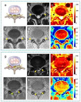 |
Screen Number: 48
0984. Ultrashort
Echo Time Magnetization Transfer Imaging of Lumbar Nerve Roots:
A Study of Low Back Pain
J. Liu, Y. Lu, Y. Ma
Ruijin Hospital, Shanghai Jiao Tong University School of Medicine, Shanghai, China
Impact: UTE-MT is a promising technique for evaluating
the compositional changes of compressed nerve roots in
lumbar radiculopathy and their relationship with LBP.
|
| 14:16 |
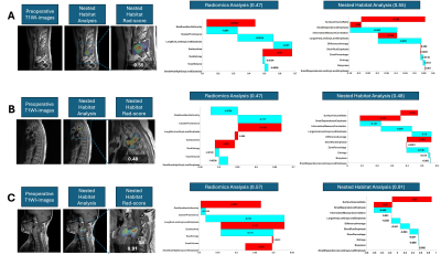 |
Screen Number: 49
0985. Nested
Habitat Analysis Based on MRI for Prediction of Progression-Free
Survival in Aggressive Spinal Tumors
Y. Zhang, Q. Wang, T. Wang, Y. Chen, R. Yan, K. Liu, W.
Zhao, D. Hao, M-Y Su, N. Lang
University of California, Irvine, United States
Impact: This study's MRI-based nested habitat radiomics
model enhances prediction accuracy of progression-free
survival (PFS) in aggressive spinal tumors by focusing on
the most aggressive regions, outperforming traditional
models and providing valuable insights for personalized
surgical and post-surgical treatment strategies.
|
| 14:18 |
 |
Screen Number: 50
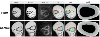
0986. Long-term
monitoring bone porosity in vivo T1DM Rabbits using UTE-MRI
k. wang, W. Liu, H. Lei, R. Yang, Y. Zha
Department of Radiology, Renmin Hospital of Wuhan University, wuhan, China
Impact: UTE-MRI is feasible to assess cortical bone
porosity and geometry in T1DM, offering a non-invasive,
radiation-free method to evaluate bone health. This
technique supports fracture evaluation and management
guideline in diabetic patients, improving clinical outcomes.
|
 Back to Meeting Home
 Back to the Program-at-a-Glance
The International Society for Magnetic Resonance in Medicine is accredited by the Accreditation Council for Continuing Medical Education to provide continuing medical education for physicians.
 View
Presentation Video
View
Presentation Video