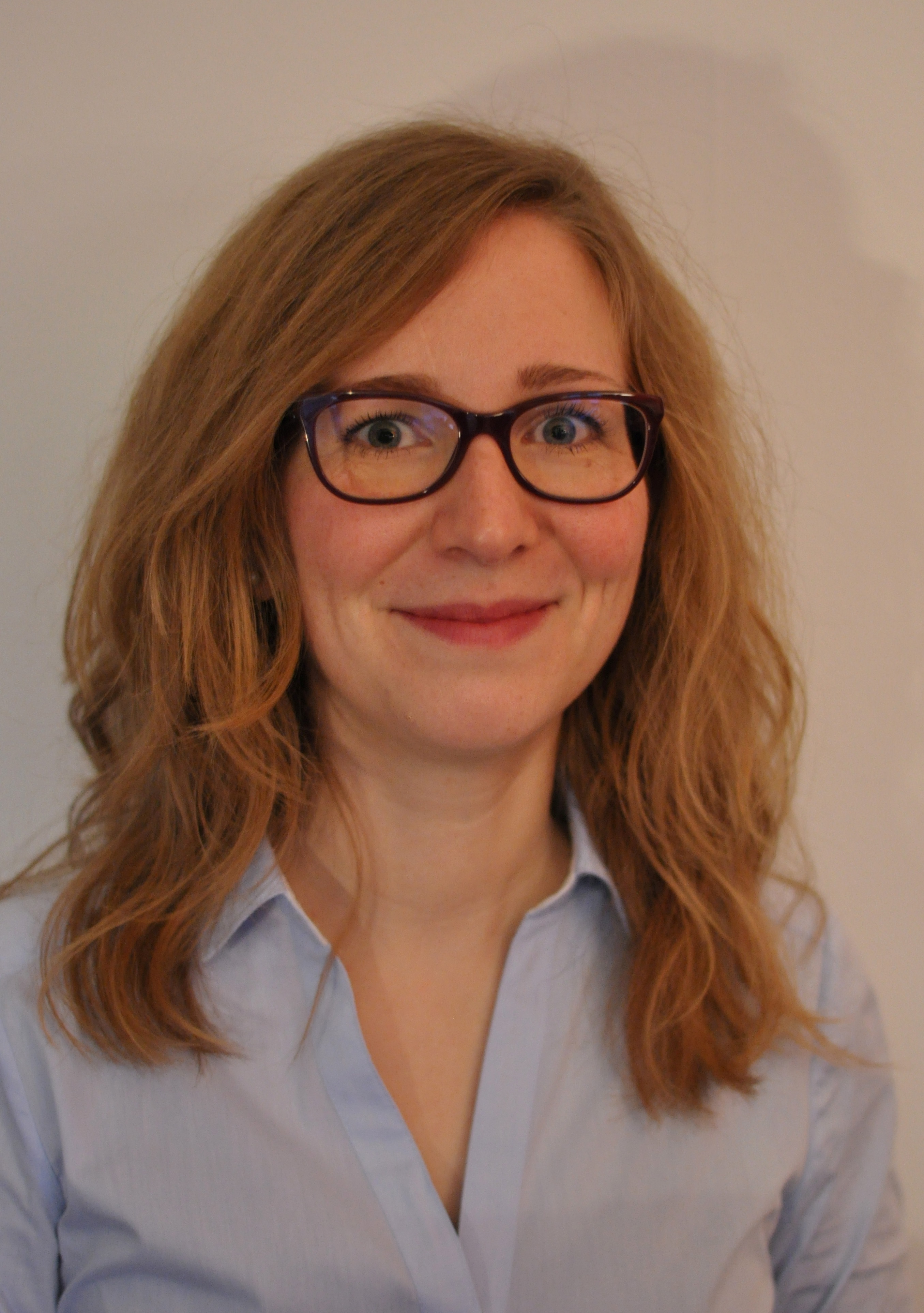

Following last week’s feature on diffusion imaging for prostate cancer characterization, this week we focus on prostate spectroscopy and a recent paper by researchers at the Radboud University Medical Center in the Netherlands, entitled ‘Improved Volume Selective 1H MR Spectroscopic Imaging of the Prostate with Gradient Offset Independent Adiabaticity Pulses at 3 Tesla’. We spoke to lead author Isabell Steinseifer and her mentor Arend Heerschap.
MRMH: Can you please tell us a bit about yourself and how you got interested in MR?
Isabell: I studied physics, and afterwards I decided to do something related to medical applications. So I came to Radboud University Medical Center, did my PhD here, and I have just started my training to become a clinical physicist in Radiotherapy.
Arend: I got a PhD at the University of Nijmegen, then moved to Philips to be involved in the development of in vivo MR. After about 5 years, I returned back to academia, and built a translational research group, with prostate MR spectroscopy as a main focus.
MRMH: Can you talk a little bit about the context, why are you interested in developing prostate MR spectroscopy and whether it is currently used in the clinic?
Isabell: We have been doing prostate spectroscopy in our group for a long time. Prostate MRSI is challenging, in particular because the prostate is small and surrounded by a lipid pool. So we developed a semi-LASER sequence with GOIA pulses, to minimize lipid contamination, and to provide more stable spectra with higher SNR. At this moment MRSI is mostly used in academic studies. Some centers are using it clinically for assessing tumor aggressiveness, and also for evaluating the effects of treatment of prostate cancer.
Arend: Prostate cancer is a very important clinical problem. MR can be important for diagnosis, but also for aggressiveness assessment, and evaluation during treatment. Over the years, we have seen the development of multi-parametric MRI, including T2 MR, diffusion MR, MR Spectroscopy, and dynamic contrast MR. Currently they come together in the so-called PI-RADS classification.
MRMH: Pirates? What are pirates?
 Arend: Not pirates, but PI-RADS, it stands for Prostate Imaging Reporting and Data System. It is a classification that radiologists use to identify the localization, and stage/grade of prostate cancer. It is important because it makes consensus diagnosis. A first version of PI-RADS included all four of the approaches listed above. Currently MR Spectroscopy and dynamic contrast are a little bit in the background. That is one of the reasons we work so hard. If you go to MR spectroscopy, you deal with much lower SNR compared to common MRI, so it is a big challenge to get a really good method in the clinic. Yet, there is ample evidence that MRS gives valuable complementary information to T2 and diffusion, so it is well worth the effort.
MRMH: What was the biggest challenge for this project?
Isabell: In this project, the most challenging part was implementing the GOIA-WURST pulses, because of the gradient modulation on top of RF modulation, which is quite difficult. But this allows us to go to low RF amplitudes, so it is worth the effort.
MRMH: MRI is dense with acronyms, but spectroscopy is the worst offender. Putting an artist (GOIA) next to a sausage (WURST) sure is memorable, but do you plan to come up with a new acronym for this sequence?
Arend: Indeed, MR is stuck with acronyms. People usually give a new acronym even if it is a small modification. We actually went the other way, we used other people’s acronyms, so for now we don’t have our own.
MRMH: What would it take to implement this at a different site?
Isabell: In the meantime our sequence has become available as a work in progress package for Siemens scanners. Going to other vendors should be possible, the important sequence parameters are described in the article. Also, we need to make sure that we are compatible with sites that do not use an endorectal coil.
MRMH: But using an endorectal coil cuts down the scan time, right?
Arend: The whole field is now moving toward removing endorectal coils. So it is important for us to demonstrate that we can run this sequence without an endorectal coil. The good news is that with the new sequence and shorter echo times we have a higher SNR, so we can also do spectroscopy without the endorectal coil.
MRMH: Patients will be happy to hear that. Where do you see this project going in the future?
Arend: We are planning clinical trials with other sites (e.g. Trondheim in Norway), using this new sequence without an endorectal coil, to demonstrate its clinical value. Our mission is to put the sequence in the clinic, so it has to be very fast. We have the ambition to shorten the spectroscopy exam to within about 7 minutes. Another challenge is post-processing. The radiologist does not want to go to a separate console to get rid of artifacts, so our ultimate ambition is to incorporate the post-processing so it all happens at the push of a button.
MRMH: We wish you success in your future endeavors!
* Interview conducted by Hong Shang and Nikola Stikov
