|
|
||||
|
July 2015 • Vol. 4, Issue 3 |
||||
| A Message from the SMRT Home Study Educational Seminars Publications Committee | ||||
 |
Anne Marie Sawyer, B.S., R.T.(R)(MR), FSMRT Home Study Program Lucas Center for Imaging Stanford University, Stanford, California, USA T: +1 650 725 9697 E: amsawyer@stanford.edu |
|||
|
MRI of the Pelvis: Prostate Cancer; Hypointense Solid Lesions; |
||||
|
We are pleased to present the SMRT Educational Seminars, Volume 18, Number 3: “MRI of the Pelvis: Prostate Cancer; Hypointense Solid Lesions; Ovarian Cystic Lesions; and Müllerian Duct Anomalies." This is the 69thaccredited home study developed by the SMRT, exclusively for SMRT members. The accreditation is conducted by the SMRT acting as a RCEEM (Recognized Continuing Education Evaluation Mechanism) for the ARRT. Category A credits are assigned to each home study, which can be used to maintain one’s ARRT advanced registry. SMRT Home Studies are also approved for AIR (Australian Institute of Radiography), NZIMRT (New Zealand Institute of Radiation Technology) and CPD Now (The College of Radiographers, United Kingdom) continuing professional development (CPD) activities.
The authors of the second article focus on T2-weighted imaging of hypointense solid lesions in the female pelvis. They introduce their strategy by saying "Malignant tissue has been shown to increase both intracellular and extracellular water, which results in increased T1 and T2 relaxation times in malignant tissue. Therefore, most solid lesions in the female pelvis appearing hyperintense on T2-weighted images should be interpreted as malignant. In contrast, solid lesions in the female pelvis that appear hypointense on T2-weighted images may be benign. Thus, distinguishing between hyperintense and hypointense solid lesions on T2-weighted images is important to help narrow the differential diagnosis and to avoid unnecessary radical surgery." In the third article the authors' discussion centers on "MRI Features of Ovarian Cystic Lesions." Despite the widely varying causes and imaging characteristics of ovarian cystic lesions, "preoperative characterization is crucial to inform women of the risks associated with radical and conservative treatment and to determine the surgical route." The article provides a wide-ranging review of cystic lesions of the ovaries. In the fourth and final article that describes Müllerian Duct Anomalies, the authors begin by reminding us that these MDA occur due to abnormal development of the uterus, cervix, and vagina, many times affecting a woman's ability to conceive and carry a pregnancy to term." The article includes a most complete review of the embryologic development of the Müllerian ducts, classification system and MRI evaluation. A special thank you to Beth Winningham, B.S.R.T., M.B.A., L.S.S.G.B. from Birmingham, Alabama, USA for acting as the Expert Reviewer. Thanks also to Heidi Berns, M.S., R.T.(R)(MR), FSMRT, Chair of the SMRT RCEEM Ad-hoc committee from Coralville, Iowa, USA and all those who participate on this committee by reviewing the home studies for accreditation. Finally, many thanks to Kerry Crockett, Associate Executive Director, Mary Keydash, Publications Director, Linda O-Brown, SMRT Coordinator, Sally Moran, Director of Electronic Communications and the entire staff in the Berkeley, California, USA office of the ISMRM and SMRT for their insight and long hours spent supporting these educational symposia. |
||||
|
Click here to continue to the SMRT Online Education website. |
||||
|
Signals is a publication produced four times per calendar year by the International Society for Magnetic Resonance in Medicine for the benefit of the SMRT membership and those individuals and organizations that support the educational programs and professional advancement of the SMRT and its members. The newsletter is the compilation of editor, Julie Strandt-Peay, BSM, RT (R)(MR) FSMRT, the leadership of the SMRT and the staff in the ISMRM Central Office with contributions from members and invited participants. |
||||

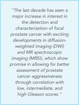 Four
previously published articles have been chosen for this home study
issue. As introduced in the first article, MRI of Prostate Cancer,
the authors begin with a historical perspective, "The advent of
rapid T2WI with fast-spin echo (FSE) techniques in the early 1990s
provided the basic tools required for prostate cancer MRI staging
examinations - fast T2WI with an endorectal coil. However, over time
the goals of clinical imaging have changed, and the entire imaging
community is shifting its emphasis towards combining
anatomical/structural imaging with functional and molecular imaging
tools." What follows is a comprehensive update of current methods
for MRI imaging of the prostate.
Four
previously published articles have been chosen for this home study
issue. As introduced in the first article, MRI of Prostate Cancer,
the authors begin with a historical perspective, "The advent of
rapid T2WI with fast-spin echo (FSE) techniques in the early 1990s
provided the basic tools required for prostate cancer MRI staging
examinations - fast T2WI with an endorectal coil. However, over time
the goals of clinical imaging have changed, and the entire imaging
community is shifting its emphasis towards combining
anatomical/structural imaging with functional and molecular imaging
tools." What follows is a comprehensive update of current methods
for MRI imaging of the prostate.