|
|||||||
| A Message from the SMRT Publications Committee about the Home Study Educational Seminars | |||||||
 |
Anne Marie Sawyer, BS RT(R)(MR) FSMRT Chair, Home Study Educational Seminars Sub-Committee |
 |
Chris Kokkinos, BAppSc PgCert MRI Chair, SMRT Online Learning Committee |
||||
|
Vol.19 #3: Body
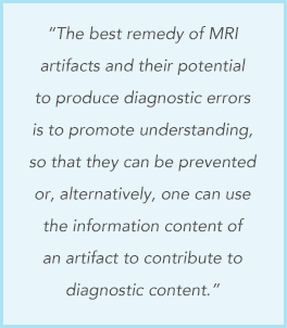
MRI Artifacts in Clinical Practice: A Physicist’s and Radiologist’s
Perspective We are pleased to present the SMRT Educational Seminars, Volume 19, Numbers 3 and 4: “Body MRI Artifacts in Clinical Practice: A Physicist’s and Radiologist’s Perspective" and “Quantification of Liver Iron with MRI: State of the Art and Remaining Challenges.” These are the 73rd and 74th accredited home studies developed by the SMRT, exclusively for SMRT members. The accreditation is conducted by the SMRT acting as a RCEEM (Recognized Continuing Education Evaluation Mechanism) for the ARRT. Category A credits are assigned to each home study, which can be used to maintain one’s ARRT advanced registry. SMRT Home Studies are also approved for AIR (Australian Institute of Radiography), NZIMRT (New Zealand Institute of Radiation Technology) and CPD Now (The College of Radiographers, United Kingdom) continuing professional development (CPD) activities.
A special thank you to Chesanie Beam, B.S., R.T.(R)(M)(MR) from Gastonia, North Carolina, USA for acting as the Expert Reviewer on both of these home studies. Thanks also to Heidi Berns, M.S., R.T.(R)(MR), FSMRT, Chair of the SMRT RCEEM Ad-hoc committee from Coralville, Iowa, USA and all those who participate on this committee by reviewing the home studies for accreditation. Finally, many thanks to Kerry Crockett, Associate Executive Director, Mary Keydash, Director of Marketing, Sally Moran, Director of IT and Web, Barbara Elliott, SMRT Coordinator, Linda O-Brown, Education Coordinator, and the entire staff in the Concord, California, USA office of the ISMRM and SMRT for their insight and long hours spent supporting these educational symposia. To access
the new Educational Seminar, |
|||||||
|
Signals is a publication produced four times per calendar year by the
International Society for Magnetic Resonance in Medicine for the benefit of the SMRT membership and those individuals and organizations that support the educational programs and professional advancement of the SMRT and its members. The newsletter is the compilation of editor, Julie Strandt-Peay, BSM, RT (R)(MR) FSMRT, the leadership of the SMRT and the staff in the ISMRM Central Office with contributions from members and invited participants.
|
|||||||

 One
peer-reviewed published article appears in each of these home study
issues. The authors of the Body Artifacts article provide a
comprehensive review of MRI artifacts encountered in imaging of the
body. “Because artifacts can arise from either the MR system
hardware alone or through the interaction of the patient with the
hardware we have chosen to present the artifacts, first, through
effects due to the static magnetic field, gradients, and
radiofrequency system, before addressing artifacts related to the
encoding of the MR signals.” Discussions of artifacts occurring in
MR imaging continue to prove useful for all radiographers and
technologists despite the length of their careers in this
technology. And hopefully many will also appreciate and be
fascinated by the underlying physics.
 The
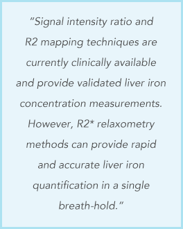
authors of the Liver Iron article offer an in-depth discussion of
iron deposition in the liver and the options to image and quantify
using MRI. “The presence of iron in tissue affects the MRI signal in
multiple ways: iron, typically in the form of ferritin and
hemosiderin, shortens the relaxation times T2*, T2 and T1 (ie,
increases the relaxation times R2*, R2 and R1). Additionally,
because iron in the body is paramagnetic, it affects the
susceptibility of tissue, resulting in macroscopic changes in the B0
field.” This most thorough review includes pathophysiology and
mechanisms of iron overload in addition to MRI-based and
non-MRI-based methods for quantification of liver iron.