ISMRT Oral
Clinical
ISMRM & ISMRT Annual Meeting & Exhibition • 03-08 June 2023 • Toronto, ON, Canada

| 19:00 |
5396.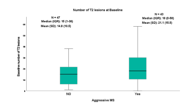 |
Predictors of long-term disability in MS patients using routine
MRI data: a 15-year retrospective study
Amjad Ibrahim Altokhis1,
Abdulmajeed Alotaibi2,
Paul Morgan2,
and Radu Tanasescu3
1Clinical Neurology, University of Nottingham, Nottingham, United Kingdom, 2University of Nottingham, Nottingham, United Kingdom, 3University of nottingham, Nottingham, United Kingdom
MRI disability predictors in Multiple Sclerosis using
routine MRI data: a 15-year longtudinal study
|
| 19:00 |
5397.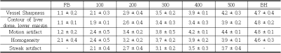 |
Comparison of 3D VANE XD Technique in T1 weighted images of
Liver MRI with free-breathing technique as the Radial percentage
parameter changes
Hun-Yeol Shin1,
Jae-Yun Jeong1,
Seong-Bong Cho1,
and Chang-Min Dae1
1Radiology, Seoul National University Bundang Hospital, Seoul, Korea, Republic of
Liver examination with MRI has greater diagnostic value than
other tests and is one of the more frequent
examinations.[1,2] However, the ability to obtain images
with good diagnostic value only when the patient breathes
constantly.[3] A non-Cartesian Radial method for the
collection of K-Space data enables examinations with free
breathing differently. 3D VANE XD technique can be applied
with mDIXON, a technique with high SNR and excellent fat
suppression.[4] Therefore, the difference in Radial
percentage, the parameter that most effects motion artifacts
in T1 3D VANE XD and 3D-FFE techniques were compared and
optimal time versus efficiency value were compared.
|
| 19:00 |
5398.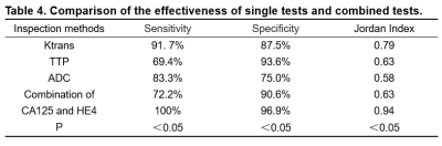 |
The application of MR perfusion and diffusion combined with
tumor marker diagnosis for the identification of benign and
malignant ovarian tumors
Chi Zhang1,
Hongying Li1,
Xin Hu1,
Jinsong Bai1,
Haitao Zhang1,
Kang Xiao1,
Kai Guo1,
and Guohua Zhang1
1HanZhong People's Hospital, Hanzhong, China
Ovarian tumors are multifaceted tumors of the female
reproductive system, of which early diagnosis and timely
treatment of ovarian tumors is particularly important. In
this study, we investigated the efficacy of dynamic
contrast-enhanced magnetic resonance imaging (DCE-MRI),
apparent diffusion coefficient (ADC), serum CA125, human
epididymal secretory protein (HE4) and their combined
application in the differential diagnosis of benign and
malignant ovarian tumors and explored the best method to be
used in the diagnosis of ovarian tumors.
|
| 19:00 |
5399.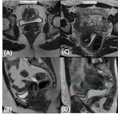 |
Improving Visualization of Cervix in MRI with Sterile Surgical
Lubricant
Stephan Jordan1,
Rebecca Rakow-Penner1,
Alex Schlein1,
Elin Lundstrom1,2,3,
Summer Batasin1,
and Stephane Loubrie4
1Radiology, UCSD, La Jolla, CA, United States, 2Department of Surgical Sciences, Uppsala University, Uppsala, Sweden, 3Center for Medical Imaging, Uppsala University Hospital, Uppsala, Sweden, 4UCSD, La Jolla, CA, United States
MRI is recommended by FIGO for staging of cervical cancer,
and thus appropriate visualization of the vaginal vault and
cervix is important for accurate staging. MRI has superior
soft tissue contrast compared to other imaging modalities.
However, when the vaginal vault is decompressed or the
prescription angle is off axis, the utility of MRI becomes
limited in evaluating cervical cancer. In this abstract, we
explore the use of sterile water-based surgical lubricant as
a tool to improve visualization of the cervix and associated
structures during MRI.
|
| 19:00 |
5400.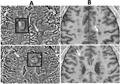 |
Longitudinal Clinical Study of Patients with Iron Rim Lesions in
Multiple Sclerosis
Amjad Ibrahim Altokhis1,2,
Aimee Hibbert1,
Christopher Allen3,
Olivier Mougin3,
Abdulmajeed Alotaibi3,
Su-Yin Lim Lim4,
Cris Constantinescu5,
Rasha Abdel-Fahim6,
Nikos Evangelou3,
and Amjad Altokhis7
1Clinical Neurology, University of Nottingham, Nottingham, United Kingdom, 2Princess Nourah bint abdulrahman University, Riyadh, Saudi Arabia, 3University of Nottingham, Nottingham, United Kingdom, 4School of Medicine, Faculty of Health and Medical Sciences, Taylor’s University, Taylor's, Malaysia, 5Department of Neurology, Cooper Neurological Institute, Cooper Neurological Institut, Camden, PA, United States, 6Queen's Medical Centre, Nottingham, United Kingdom, 7Clinical Neurology, University of nottingham, Nottingham, United Kingdom
Iron rim lesions in Multiple Sclerosis disability
|
| 19:00 |
5401.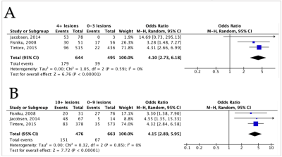 |
Magnetic Resonance Imaging as a Prognostic Disability biomarker
in C Multiple Sclerosis: A Systematic Review and Meta-Analysis
Amjad Ibrahim Altokhis1,
Abrar Alamrani2,
Abdulmajeed Alotaibi3,
Anna Podlasek4,
and Amjad Altokhis5
1Clinical Neurology, University of Nottingham, Nottingham, United Kingdom, 2Faculty of Health, York University, Toronto, ON, Canada, 3University of Nottingham, Nottingham, United Kingdom, 4anna.podlasek@nottingham.ac.uk, Nottingham, United Kingdom, 5Clinical Neurology, University of nottingham, Nottingham, United Kingdom
Imaging biomarker for disability in Multiple Sclerosis
|
| 19:00 |
5402.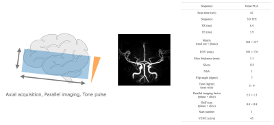 |
The Imaging Technique and Clinical Usefulness of Super Rapid
Phase Contrast Angiography for Stroke Patients.
Daisuke Oura1,
Riku Ihara2,
Takumi Yokohama2,
Yoshimasa Niiya3,
Koji Furukawa3,
Masayuki Gekka3,
and Hiroyuki Sugimori4
1Graduate School of Health Sciences, Hokkaido University, Sapporo, Japan, 2Radiology, Otaru General Hospital, Otaru, Japan, 3Neurosurgery, Otaru General Hospital, Otaru, Japan, 4Faculty of Health Sciences, Hokkaido University, Sapporo, Japan
We offer super rapid phase contrast angiography (PCA) with a
60-second scan. This sequence greatly contributes to
managing stroke patients compared with a conventional
technique such as time-of-flight MRA. Parallel imaging and
optimized voxel size reduce scan time as possible. Tilted
optimized non-saturating excitation enhanced the depiction
of the distal arteries in transverse acquisition PCA. Super
rapid PCA overcomes motion artifacts due to short scan time
and the shortest repetition time such as under 7 ms.
Moreover, we obtain both MRA and black blood images as a
subtraction image between the magnitude image and the MRA
image.
|
| 19:00 |
5403.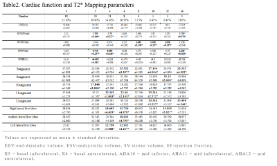 |
Incremental value of right ventricle function and T2* Mapping
for judging the occurrence and development of cirrhotic
cardiomyopathy in Rabbits
Xinai Zhang1,
Wanyin Qi1,
Zhengyuan Xiao1,
Xiaoyong Zhang2,
and Jing Chen1
1The Affiliated Hospital of Southwest Medical University, Luzhou, China, 2Philips Healthcare, Chengdu, China
This study analyzed the T2* mapping sequence and ventricular
function parameters to assess cardiac injury in early
cirrhotic cardiomyopathy (CCM) in a rabbit model. At
the 2ed week, the ejection fraction (EF) of right ventricle
(RV) decreased and the T2* value of 10th segment for LV
increased significantly. The liver showed mild iron
deposition at the 4th. T2* value of the 10th segment was
negatively correlated with that of liver. Suggesting the
iron deposition in CCM were not synchronous. Finding the T2*
mapping combine right ventricle function for evaluating the
occurrence and development of cardiac and liver
injury with CCM.
|
| 19:00 |
5404.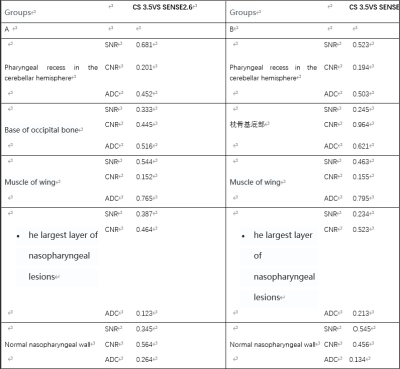 |
Application of diffusion-weighted imaging of fast spin echo
sequences based on compressed sensing in nasopharyngeal imaging
wang shuai1
1Department of Diagnostic Radiology, Xijing Hospital, Air Force Medical University, Xi'an, China
There are two imaging options for nasopharyngeal DWI, one is
EPI self-selected echo sequence, and the other is fast spin
echo sequence. The two sequences are completely different in
the way of reading signals, and each has its own advantages
and disadvantages. Traditional EPI imaging has high
distortion, and its advantages are fast scanning speed,
while TSE sequence has small deformation, but it takes a
long time. It takes nearly 4 minutes to scan a complete
nasopharynx. In view of the long scanning time and
distortion, we put forward two questions. The first question
is how to shorten the imaging time while ensuring the image
quality; the second question is what is the optimal
compression factor for nasopharynx?
|
| 19:15 |
5405.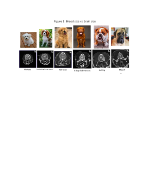 |
Is Veterinary MRI feasible in a human facility?
Shiami Delina Luchow1 and
Saad Ramadan2
1MRI, Hunter Medical Research Institute/University of Newcastle, New Lambton Heights, Australia, 2HMRI Imaging Centre, Hunter Medical Research Institute/University of Newcastle, New Lambton Heights, Australia
Magnetic resonance imaging is continuing to grow for
clinical diagnosis in veterinary practice. Although MRI is
the gold standard for imaging the central nervous system and
musculoskeletal pathology in animals, the use of MRI is
limited due to the difficulty in accessing and the higher
running cost. It is a limited resource disadvantaging many
vet animals and their owners of care and cost. Is it
feasible for a human MRI facility to image animals? This
paper discusses how this was achieved at the Hunter Medical
Research Institute Imaging Centre, Newcastle, Australia, and
the safety procedures that were necessary for approval.
|
| 19:15 |
5406.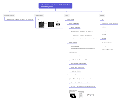 |
Radomics Model and Deep Learning Model Based on T1WI Image for
Acute Lymphocytic Leukemia Identification
Ting Yi1,
Hui Tang2,
Yuanbin Chen2,
Qifang Cai1,
Huiting Zhang3,
Weian Wei1,
and Ke Jin1
1Hunan Children's Hospital, Changsha, China, 2Fuzhou University, Fuzhou, China, 3MR Scientific Marketing, Siemens Healthineers, Guangzhou, China
This study investigated the feasibility of radomics model
and deep learning model Based on T1WI image for acute
lymphocytic leukemia identification. The results showed that
both radomics model and deep learning model can effectively
distinct ALL children and normal children. And radomic model
is better.
|
| 19:15 |
5407.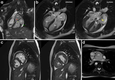 |
Cardiovascular magnetic resonance in the evaluation of
congenitally corrected transposition of the great arteries
Jose Ngombo-Kimbongila 1,
Petronella Samuels2,
Morne Kahts2,
Stephen Jermy2,
Sulaiman Moosa3,
Blanche Cupido4,
and Ntobeko Ntusi4,5
1Department of Radiology, Groote Schuur Hospital, Cape Town, South Africa, 2Cape Universities Body Imaging Centre, University of Cape Town, Cape Town, South Africa, 3Department of Radiation Medicine, University of Cape Town and Groote Schuur Hospital, Cape Town, South Africa, 4Division of Cardiology, Department of Medicine, University of Cape Town, Cape Town, South Africa, 5Cape Heart Institute, Faculty of Health Sciences, University of Cape Town, Cape Town, South Africa Congenitally corrected transposition of the great arteries (ccTGA), is a rare cardiac anomaly (occurring in less than 1% of all congenital heart diseases) characterised by atrio-ventricular and ventricular-arterial discordance. We report on a 13-year-old female with situs inversus totalis, ccTGA, peri-membranous VSD, and sub-PS presenting with worsening fatigue on physical exertion, excessive sweating, and intermittent palpitations not associated with physical activity. Echocardiography was used in diagnosis. CMR revealed mild systolic impairment (ejection fraction, EF 55%) of the systemic ventricle (morphologic RV), a perimembranous VSD, flattening of the interventricular septum in systole and diastole, indicative of pressure and volume overload, respectively.
|
| 19:15 |
5408.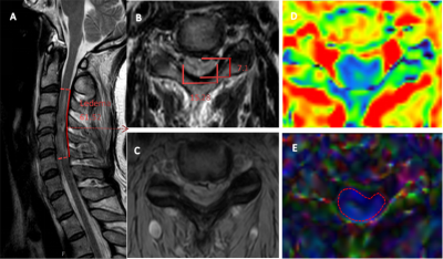 |
Evaluation the intramedullary severity and prognosis of early
MRI in adult cervical spinal cord injury without radiologic
abnormalities
Yuan Liu1,
Peng Sun2,
and Xiangchuang Kong1
1Department of Radiology, Union Hospital, Tongji Medical College, Huazhong University of Science and Technology, Wuhan, China, 2Philips Healthcare, Beijing, China The mpMRI biomarkers could be insightful for pathogenesis and prognosis in early-stage SCIWORA. |
| 19:15 | 5409. |
Application of T1 radial vibe sequence in fetal central nervous
system
TIAN JIAN1
1XiJing Hospital, XI'AN, China
Application of T1 radial vibe sequence in fetal central
nervous system
|
| 19:15 |
5410.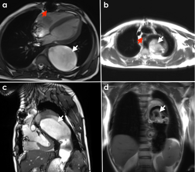 |
Phenotypic characterisation of multifocal cardiovascular
involvement in Takayasu arteritis with cardiovascular magnetic
resonance
Mariaan Jaftha1,
Petronella Samuels2,
Morne Kahts1,
Stephen Jermy2,
Tasnim Bana3,
and Ntobeko Ntusi1,4,5
1Cape University Body Imaging Centre, University Cape Town, Cape Town, South Africa, 2Cape University Body Imaging Centre, CUBIC University Cape Town, Cape Town, South Africa, 3Division of Cardiology,Department of Medicine,University of Cape Town, University of Cape Town, Cape Town, South Africa, 4Division of Cardiology,Department of Medicine,University of Cape Town, University Cape Town, Cape Town, South Africa, 5Cape Heart Institute,Faculty of Health Sciences, University Cape Town, Cape Town, South Africa
Takayasu arteritis (TA) is an uncommon inflammatory disease
primarily affecting the aorta and its main branches. It is
more common in females (80-90% of cases) and occurs between
the ages of 10 and 40 years. We report on a young male
patient diagnosed with TA at age 16 years. CMR showed
progressive aneurysmal dilatation of the aorta compressing
the trachea, left main bronchus, and left lung. Both
ventricles showed mild decrease in function (LVEF- 45% and
RVEF – 51%). CMR played an important role in disease
monitoring and guided patient management.
|
| 19:15 |
5411.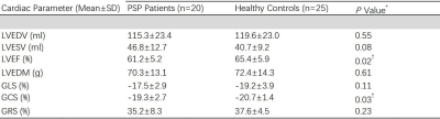 |
The potential role of FT - CMR for detecting left ventricular
dysfunction in patients with PSP: a case control study
Pengfei Peng1,
Xun Yue1,
jia yu sun1,
and Pengfei Peng1
1Radiology, West China Hospital, Sichuan University,, Chengdu, China
The potential role of feature tracking - CMR for detecting
left ventricular dysfunction in patients with PSP
|
| 19:15 |
5412. |
Dog breed size versus brain size and its inferences, and canine
pathology case studies
Shiami Delina Luchow1
1MRI, Hunter Medical Research Institute/University of Newcastle, New Lambton Heights, Australia
Colloquially intelligence is correlated to brain size.
Although not accurate in humans, is this true in dogs? Dogs
are diverse in shapes and sizes and are bred for their
unique abilities and behavioural characteristics. However,
during clinical MRI examinations, it was clear that although
the body mass ranged from approximately 1kg to over 100kg in
different breeds, the brain size did not vary drastically.
This study will analyse the neurocephalic index of different
breeds of dogs to evaluate if this correlates with the dog
breeds’ unique abilities.
|
| 19:15 | 5413. |
The conspicuity of inner ear membranous labyrinth anatomy using
3D FLAIR without gadolinium contrast agent
Zongrui Zhang1,
Zhaohui Liu1,
and Yantao Niu1
1Radiology, Beijing Tongren Hospital,Capital Medical University, Beijing, China
Optimized 3D-FLAIR imaging can visualize inner ear
membranous labyrinth anatomy without gadolinium contrast
agent,paving the way toward developing a simple and quick
method for diagnosing Meniere’s disease.
|
| 19:30 |
5414.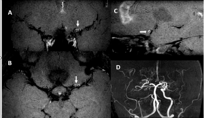 |
Role of Intracranial Vessel Wall (VW) MRI in Evaluating Luminal
Pathologies.
Hui Ping Oh1,
Ei Nyein Ei2,
and Soke Miang Chng2
1Neuroradiology, National Neuroscience Institute, Singapore, Singapore, 2National Neuroscience Institute, Singapore, Singapore
Intracranial vessel wall (VW) MRI is a state-of-art
technology to evaluate vessel wall diseases. It requires
high spatial and contrast-to-noise ratio (CNR) resolution as
well as capability of blood and cerebrospinal fluid (CSF)
suppression to visualize the arterial wall. Our vessel wall
protocol including T2 -weighted, 3D time of flight (ToF) MR
angiography, diffusion-weighted imaging (DWI), gradient echo
imaging (GRE), high resolution post contrast 3D T1-weighted
turbo spin echo (TSE) with motion-sensitized
driven-equilibrium (MSDE) black blood sequence and post
contrast 3D T1-weighted volumetric isotropic fast field echo
(FFE) done within 30 minutes which is capable to show vessel
wall diseases.
|
| 19:30 |
5415.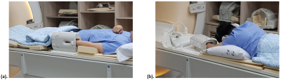 |
Comparative evaluation of fiber number implementation of median
nerve during wrist DTI technique : Neutral vs Superman position
Seong-Bong Cho1 and
Jae-Yun Jeong1
1radiology, Seoul National University Bundang hospital, Gyeonggi-do, Korea, Republic of
To find out whether this change in position affects the
actual fiber tracking, we compared the number of fiber
implementations in the median nerve in accordance to neutral
and superman position. In 12 of the 14 cases, the superman
position produced more fibers than in the neutral
position. The reason for this thought is due to the fact
that the wrist in the superman position is closer to the
isocenter of the magnetic field. Other parameters may be
important to optimize the images of wrist DTI, however
positioning of the wrist should be considered first.
|
 Back to Meeting Home
 Back to the Program-at-a-Glance
The International Society for Magnetic Resonance in Medicine is accredited by the Accreditation Council for Continuing Medical Education to provide continuing medical education for physicians.
 Back to Meeting Home
 Back to the Program-at-a-Glance
 View Presentation Video