ISMRT Oral
Research
ISMRM & ISMRT Annual Meeting & Exhibition • 03-08 June 2023 • Toronto, ON, Canada

| 19:30 |
5416.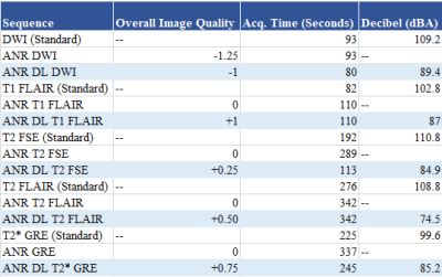 |
Acoustic Noise Reduction in MRI and Utilizing Machine Learning
Ian David Langenfeld RT.(R)(MR)(MRSO)1,
Jenna Kleinow RT.(R)(MR)(MRSO)1,
Brian Burkett M.D., M.P.H.1,
Paul Farnsworth D.O.1,
Garima Suman, M.D. 1,
Steven Messina, M.D.1,
and Joel Felmlee Ph.D.1
1Radiology, Mayo Clinic, Rochester, MN, United States
This study assesses the use of acoustic noise reduction (ANR)
techniques as well as utilizing machine learning in
conjunction with ANR, in order to preserve image quality and
limit acquisition time. The standard sequences in a brain
without contrast exam were tested to determine decibel (dBA)
level, image quality, as well as the scanned volunteers'
semiquantitative "loudness" scores.
|
| 19:30 |
5417.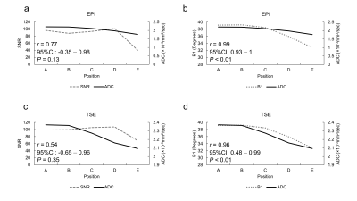 |
The position dependence of the apparent diffusion coefficient;
signal-to-noise ratio vs B1 map
YASUO TAKATSU1,2,
Masafumi Nakamura2,3,
Yuichi Suzuki4,
and Tosiaki Miyati2
1Molecular Imaging, School of Medical Sciences, Fujita Health University, Toyoake, Japan, 2Division of Health Sciences, Graduate School of Medical Sciences, Kanazawa University, Kanazawa, Japan, 3Department of Radiology, Otsu City Hospital, Otsu, Japan, 4Department of Radiology, The University of Tokyo Hospital, Tokyo, Japan
The position dependence of the ADC in magnetic resonance
imaging (MRI) by EPI- and TSE-DWI was assessed using
phantoms based on the relationship between SNR and B1 maps.
ADC decreased with distance from the center of the magnetic
field for both EPI-DWI and TSE-DWI. The Pearson correlation
coefficient between ADC and FA was strong and more
significant than between ADC and SNR. ADC depends on the
slice position and decreases with an increase in distance
from the magnetic field center. Caution should be taken when
comparing and quantitatively evaluating the ADC at sites
shifted in the long-axis direction.
|
| 19:30 |
5418.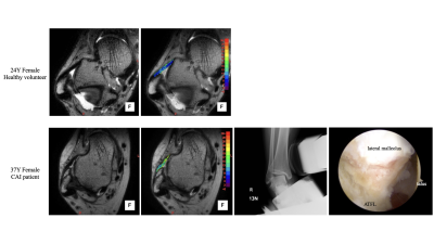 |
Quantitative assessment of anterior talofibular ligament quality
in chronic ankle instability using T2* relaxation time
Yoshihiro Akatsuka1,
Atsushi Teramoto2,
Yasutaka Murahashi2,
Katsunori Takahashi2,
Rui Imamura1,
Tomoaki Kamiya2,
and Kota Watanabe3
1Division of Radiology and Nuclear Medicine, Sapporo Medical University Hospital, Sapporo, Japan, 2Department of Orthopedic Surgery, Sapporo Medical University School of Medicine, Sapporo, Japan, 3Second Division of Physical Therapy, Sapporo Medical University School of Health Sciences, Sapporo, Japan
This study investigated the feasibility of quantitative
assessment of anterior talofibular ligament (ATFL) quality
in chronic ankle instability (CAI) using T2* relaxation
time. A prediction equation for the normal ATFL reference
value was calculated in healthy volunteers. The ratio
between the normal reference value and the patient's T2*
value showed a significant positive correlation with the
talar tilt angle on stress radiograph. There was also
increasing trend in T2* values for poor ligament quality in
the arthroscopic findings. T2* relaxation times are
promising for quantitatively assessing ATFL quality
preoperatively.
|
| 19:30 |
5419.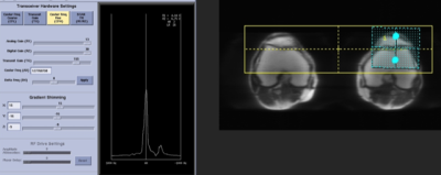 |
Comparing Dual Shimming with Average Single Shimming on Fat
Suppression Techniques
Vahid Ravanfar1,
Heather Daniel1,
Emma Bahrros1,
Rupsa Bhattacharjee1,
Maya Aslam1,
Patrick D. Koon2,
Mehdi Khalighi3,
and Dr. Matthew Bucknor1
1Radiology and Biomedical Imaging, University of California San Francisco, San Francisco, CA, United States, 2GE Healthcare, Waukesha, WI, United States, 3Rad/PET/MRI Metabolic Service Center, Stanford University, Palo Alto, CA, United States
When acquiring fat suppression sequences, active shimming
can be used to reduce magnetic field inhomogeneities which
are inversely related to image quality and cause image
artifact. Active shimming improves image quality by
optimizing the homogeneity on an individual patient basis to
optimize the final shimming and directly, the resulting
image quality.
|
| 19:30 |
5420.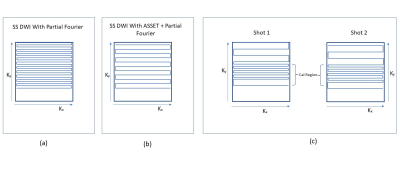 |
DWI of liver using combination of sub-sampling and dual shots
without dedicated multi-channel abdomen coil- A CT like patient
positioning
Sajith Rajamani1,
Ashok Kumar Reddy1,
Nitin Jain1,
Rajdeep Das1,
Rajagopalan Sundaresan1,
Jeremy Heinlein2,
Harsh Kumar Agarwal1,
Arnaud Guidon2,
Sudhir Ramanna1,
and Ramesh Venkatesan1
1GE Healthcare, Bangalore, India, 2GE Healthcare, Milwauke, WI, United States
DWI is an important sequence for the diagnosis of liver
lesions and to monitor the treatment response in patients
undergoing therapies for hepatic malignancies [1]. But it is
difficult to place the anterior abdomen coil for obese
patients in non-wide bore MRI systems because of a potential
chance of pinching the coil between patient and the bore.
This is a limiting factor for the adoption of liver DWI MRI
in clinical practice [2]. As a solution, we are proposing
Dual Shots DWI with under-sampling across shots using volume
coil located inside the magnet.
|
| 19:30 |
5421.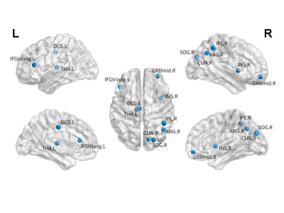 |
Characteristics of brain white matter network in adolescent
patients with first-episode non-suicidal self-injury
Yuwei Chen1 and
Nian Liu1
1North Sichuan Medical College, Nanchong, China Objective: Explore the characteristics of brain white matter network in non-suicidal self-injury patients. Methods: The diffusion tensor imaging were prospectively collected form 30 adolescent patients with NSSI. The data was processed by software. Two independent sample t-test and partial correlation analysis were used to compare network metrics and clinical symptoms. Results: There was no significant difference in global network metrics, but there were significant differences in nodal network metrics, including nodal efficiency, nodal degree centrality and nodal shortest path length (all P<0.05). Conclusion: These finding provide new insights into the neural circuit mechanism of adolescent NSSI patients. |
| 19:30 |
5422.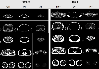 |
Correlation between body fat distribution characteristics and
human body parameters based on magnetic resonance imaging
YuLong Qi1,
GuanXun Cheng2,
and ChuanLi Cheng3
1Medical Imaging Department, Peking University ShenZhen Hospital, ShenZhen, China, 2Peking University ShenZhen Hospital, ShenZhen, China, 3Shenzhen Institutes of Advanced Technology(SIAT), ShenZhen, China
The study explores the correlation between fat distribution
characteristics and traditional anthropometric indexes.37
volunteers were scanned with whole-body transverse MR
PDFF(Proton Density Fat Fraction) images, covering from neck
to knee.In the fat distribution characteristics based on
magnetic resonance whole-body imaging, only the proportion
of whole-body fat volume showed moderate correlation with
traditional ergonomic indexes, and other indexes had low
correlation with ergonomic indexes.the correlation between
the whole-body fat distribution characteristics based on
PDFF image and body mass index (BMI) ,waist to hip ratio
(WHR) was analyzed. Different from the anthropometric
indexes, the distribution characteristics of body fat show
great differences between sexes.
|
| 19:45 |
5423.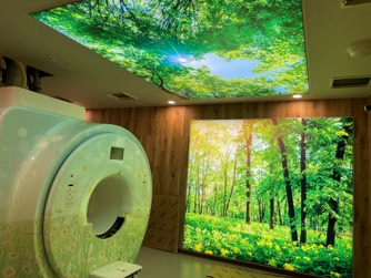 |
Investigation of mental state of patients after an examination
in an MRI room with LED-backlight photoprints
Hiroyuki Hoshiko1,
Masaaki Ninomiya1,
Akiyoshi Yamamoto1,
Seigo Yoshida1,
and Katsumi Nakamura1
1Tobata Kyouritsu Hospital, Kitakyusyu,Fukuoka, Japan
We investigated the psychological state of patients in an
MRI examination room equipped with LED-backlit photoprints
(SKY LIGHT). We surveyed patients who had completed their
examinations in the room about their impressions of the
room. The patients' responses were categorized into three
types based on their words: positive, negative, and
indifferent, and the percentages were calculated. 74.5% of
the patients had positive impressions of the rooms after the
examination. In conclusion, the results suggest that MRI
rooms with SKY LIGHT can give positive impressions to
patients.
|
| 19:45 | 5424. |
Incidence of Meniscal tears associated with osteoarthritis on
MRI Knee joint: MMI Hospital Karachi.
ABDUL QAYOOM RAKHSHANI1 and
Uzama Azmat2
1RADIOLOGY۔ MRI, DOW UNIVERSITY OF HEALTH SCIENCES KARACHI, Karachi, Pakistan, 2Radiology, MMI Karachi, Karachi, Pakistan To assess the yield of Incidence of Meniscal tears and associated with osteoarthritis on MRI Knee joint non-contrast enhanced. A cross Sectional study was conducted at department of Radiology MRI Section MMI HOSPITAL Karachi. From January 2019 to February 2020. All patients came for MRI Examination; we assess MRI Knee Joints Non-Contrast Enhanced for Association of meniscal tears with Osteoarthritis on Knee Joints. The data was analyzed for demographic characteristics, referring clinician and Site, Grade and type of tears with final diagnosis. |
| 19:45 |
5425.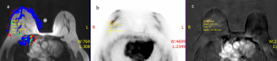 |
Metabolic Tumor Volume prediction by using Hand Craft Fuzzy Rule
Base System (FRBS) on Simultaneous PET/MRI
Pradeep Singh Negi1,2,
Shashi Bhushan Mehta1,2,
and Amarnath Jena1,2
1Department of Molecular Imaging and Nuclear Medicine, PET SUITE: Indraprastha Apollo Hospitals and House of Diagnostics, Delhi, India, 2Department of Physics, Vivekananda Global University, Jaipur, India 18F-FDG PET/MRI can be helpful for diagnosing, staging, restaging, and assessing the post therapeutic response in breast cancer patients. Metabolic tumor volume (MTV) may be more reliable but volumetric assessments of FDG PET needs correct tumor segmentation. We have proposed hand craft fuzzy rule base system to improve tumor volume by using simultaneous PET/MRI for accurate MTV estimation. Four breast cancer patients underwent FDG PET/MRI for staging purpose. Pathological tumor volume was compared with MRI volume, Ktrans volume and MTV computed by hand craft fuzzy rule and the results showed fuzzified tumor volume were more accurate compared with other volumes. |
| 19:45 | 5426. |
Clinical feasibility study of AI accelerateed STAGE
Yang Sun1
1First Hospital of Jilin University, ChangChun, China
Clinical feasibility study of AI accelerateed STAGE
|
| 19:45 |
5427. |
Reducing contrast agents’ residuals in hospital wastewater: the
GREENWATER study
Moreno Zanardo1,
Luigi Asmundo1,
Davide Capra2,
Anna Colarieti3,
Andrea Cozzi4,
Massimo Cressoni4,
Veronica Magni1,
Caterina Beatrice Monti2,
and Francesco Sardanelli5
1Università degli Studi di Milano, Milano, Italy, 2Università degli Studi di Milano, Milan, Italy, 3IRCCS Policliico San Donato, Milano, Italy, 4IRCCS Policlinico San Donato, Milano, Italy, 5Università degli Studi di Milano - IRCCS Policlinico San Donato, Milan, Italy
In order to provide preliminary data about the potential
reduction of contrast agents’ residuals in hospital
wastewater and to estimate contrast agent excretion in the
first hour after administration, the GREENWATER study aims
to prospectively monitor over a 12-months timeframe the
quantity of retrievable ICAs and GBCAs from urine collected
from outpatients within an hour from contrast agent
administration, also evaluating the influence of patient age
and sex and the overall rate of acceptance to participate to
the study. Our current purpose is to provide a first glance
on this initial experience.
|
| 19:45 |
5428.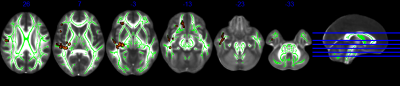 |
The Quantitative Evaluation of Mild Traumatic Brain Injury with
DSI and DTI MR Technology
JinRui Zhang1
1Chongqing Emergency Medical Center, Chongqing, China
Compared with DTI (diffusion tensor imaging), DSI(diffusion
spectrum imaging) has more accurate spatial resolution
ability in mTBI(mild traumatic brain injury), and DSI-based
fiber tracking technology has become an important tool for
medium scale (mesocale) structural elucidation, building a
bridge between microscopic and macroscopic scales, which
provides the possibility for further exploration and
integration of multi-scale analytical studies at the
cellular level as well as at the subcellular level.
|
 Back to Meeting Home
 Back to the Program-at-a-Glance
The International Society for Magnetic Resonance in Medicine is accredited by the Accreditation Council for Continuing Medical Education to provide continuing medical education for physicians.
 Back to Meeting Home
 Back to the Program-at-a-Glance
 View Presentation Video