ISMRT Oral
1st, 2nd & 3rd Place Research & Clinical Poster Winners
ISMRM & ISMRT Annual Meeting & Exhibition • 03-08 June 2023 • Toronto, ON, Canada

ISMRT Oral
1st, 2nd & 3rd Place Research & Clinical Poster Winners
| 18:00 |
5390.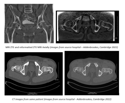 |
The benefits of ZTE to standard MRI practice
Helen L Prince1
1MRI, CUH Addenbrookes UK, Newmarket, United Kingdom
In theory a ZTE or Zero Echo Time could visualise and
benefit MRI imaging of any joint. It can be used in a wide
spectrum of developmental, traumatic, inflammatory,
rheumatologic and oncologic conditions. It may remove the
need for CT with detailed depiction of bone anatomy. It
opens the doors for more MRI based research into many
musculoskeletal conditions and morphometric analysis. One
MRI examination with a ZTE sequence allows cross referencing
of sequences aiding diagnosis, prognostication and surgical
guidance in soft tissue and bone with precise measurements
that involve bony landmarks.
|
| 18:00 |
5391.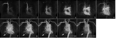 |
Clinical Impact of Single Breath Hold Contrast Enhanced 4D-MRA
with High Temporal and Spatial Resolution without k-space Data
Sharing Techniques
Tatsunori Saho1,
Johshin Matsuzaki1,
Chihiro Hayashida1,
and Takahiro Kubota1
1Dept. of Radiological technology, Kokura memorial hospital, Kitakyushu, Japan
For evaluation of arteriovenous malformations,
contrast-enhanced 4D-MRA is useful to detect feeding
arteries and draining veins. However, with the Keyhole
technique, contamination of different phases is a major
problem. We solved this problem by applying parallel imaging
with compressed sensing to contrast-enhanced
4D-MRA(CS-4D-MRA). CS-4D-MRA was able to acquire images
without contamination of the arterial phase with echo
signals from the venous phase. It also had high temporal and
spatial resolution, and was able to clearly visualize the
feeder and the drainer.
|
| 18:00 |
5392.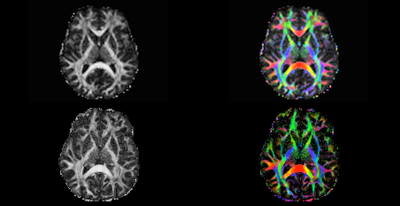 |
Diffusion imaging: multi-shell DTI on a whole-body 3T scanner
versus a head-only MAGNUS 3T for traumatic brain injury
evaluation
Gail H Kohls1,2,
Herman Douglas Morris1,2,
Maureen N Hood1,2,
James Kevin DeMarco1,2,
and Thomas KF Foo1,3
1Radiology & Radiological Sciences, USUHS, Bethesda, MD, United States, 2Radiology, Walter Reed National Military Medical Center, Bethesda, MD, United States, 3GE Research Center, Niskayuna, NY, United States Diffusion imaging has progressed beyond standard DTI to mutli-shell and non-Gaussian techniques to improve upon the sensitivity of detecting multiple fiber angles in a voxel. Newer high-gradient scanners are able to further expand the capabilities of these advanced DTI sequences to help us improve upon the detection of complex fiber tracks in voxels, which is important in the evaluation and treatment of traumatic brain injury. These new technologies hold promise to improve our understanding of the movement of microcellular fluids. |
| 18:00 |
5393.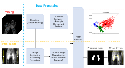 |
RA synovitis segmentation based on unsupervised learning and TIC
signal data on DCE-MRI
YiJun Mao1,2,
Wanxuan Fang2,
Yujie An2,
Hiroyuki Sugimori1,
Shinji Kiuch3,
and Tamotsu Kamishima1
1Faculty of Health Sciences, Hokkaido University, Sapporo, Japan, Sapporo, Japan, 2Graduate School of Health Sciences, Hokkaido University, Sapporo, Japan, Sapporo, Japan, 3AIC Yaesu Clinic, Tokyo, Japan, Tokyo, Japan Keywords: Rheumatoid Arthritis, DSC & DCE Perfusion The volume of synovitis change is one of the most important pathological features of rheumatoid arthritis. By quantitative analysis of the enhancement of synovitis, we can define the degree of the disease, and determine the treatment and diagnosis. Considering the time-consuming of manual outlining and visual assessment, this study uses machine learning methods to conduct quantitative analysis of TIC, and proposes an unsupervised learning method with excellent results, which is expected to be an alternative for the gold-standard manual synovitis contour outlining. |
| 18:00 |
5394.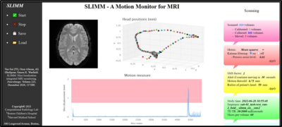 |
Technologist Assessment of a Realtime Motion Monitoring System
for fMRI Exams
Kristina M. Pelkola1,2,
Norman Farrar1,
Alyssa Ailion3,
Tess Wallace1,2,
Onur Afacan1,2,
Yao Sui1,2,
and Simon K. Warfield1,2
1Radiology, Boston Children's Hospital, Boston, MA, United States, 2Computational Radiology Laboratory, Boston Children's Hospital, Boston, MA, United States, 3Neurology, Boston Children's Hospital, Boston, MA, United States
Visit any magnetic resonance imaging (MRI) facility, and all
will agree that patient motion is an ongoing matter. This
holds particularly true for pediatric facilities performing
functional MRI (fMRI) exams. During fMRI exams, motion
artifacts are subtle and contribute to data corruption which
is not discovered until the data is analyzed. To enable the
technologist to intervene when motion occurs, a real-time
motion monitoring system “Slice Localization Integrated MRI
Monitoring” (SLIMM) was established to detect motion and
reduce the amount of scan time necessary while
simultaneously increasing the quality of data collected.
|
| 18:00 |
5395.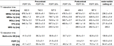 |
Evaluation of T1 relaxation time measurement using magnetic
resonance spectroscopy unobstructed by the presence of fat: A
liver phantom study.
Makoto Suzuki1,
Tatsyuya Hayashi2,
Kazutaka Nashiki1,
Hidemichi Kawata1,
Shuji Nagata3,
and Toshi Abe3
1Department of Radiological technology, Kurume University Hospital, Kurume, Japan, 2Department of Radiological Technology, Faculty of Medical Technology, Teikyo University, Tokyo, Japan, 3Department of Radiology, Kurume University School of Medicine, Kurume, Japan
We investigated the usefulness of magnetic resonance
spectroscopy (MRS) for water T1 relaxation
time independent on the presence of fat in the liver
phantom. Then,T1 relaxation
time measurements were performed using inversion
recovery-spin echo, modified look locker, variable flip
angle, and MRS on a 3T-MRI system. T1 relaxation
time measurement by the MRS water signal is less affected by
the presence of fat and more accurate than the other
methods. This technique does not use special research
sequences and can be realized on clinical MRI scanner where
MRS can be performed.
|
 Back to Meeting Home
 Back to the Program-at-a-Glance
The International Society for Magnetic Resonance in Medicine is accredited by the Accreditation Council for Continuing Medical Education to provide continuing medical education for physicians.
 Back to Meeting Home
 Back to the Program-at-a-Glance
 View Presentation Video