Digital Poster
Modeling II
Joint Annual Meeting ISMRM-ESMRMB & ISMRT 31st Annual Meeting • 07-12 May 2022 • London, UK

| Computer # | ||||
|---|---|---|---|---|
1497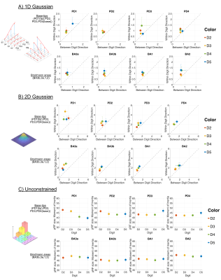 |
47 | Two-dimensional population receptive field mapping of human primary somatosensory cortex
Michael Asghar1, Rosa Sanchez Panchuelo2, Denis Schluppeck1, and Susan Francis1
1University of Nottingham, Nottingham, United Kingdom, 2University Hospitals Birmingham NHS Foundation Trust, Birmingham, United Kingdom
High-resolution fMRI data of somatosensory cortex were obtained at 7T (n=7 participants) using a 4x4 grid of piezoelectric stimulators. Population receptive field (pRF) mapping was performed using different model specifications (1D Gaussian within-digit, 1D Gaussian between-digit, 2D Gaussian of within-digit and between-digit, Unconstrained model) and pRF sizes were estimated. Grouping pRF response by 4 preferred locations within digits (along tip-base axis) or by Brodmann areas revealed more elliptical pRFs within digit representations of D2 to D4 and more circularly symmetric pRFs for D5. We also found relatively larger pRF sizes in D5 and towards the base of the digits.
|
||
1498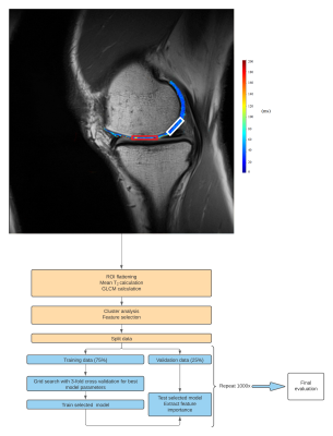 |
48 | GLCM texture analysis of knee cartilage T2 maps: machine learning based selection of important features
Veronika Janacova1, Pavol Szomolanyi1, Dominik Vilimek1,2, Siegfried Trattnig1,3,4, and Vladimir Juras1
1High Field MR Centre, Department of Biomedical Imaging and Image-Guided Therapy, Medical University of Vienna, Vienna, Austria, 2Department of Cybernetics and Biomedical Engineering, VSB–Technical University of Ostrava, Ostrava, Czech Republic, 3CD laboratory for Clinical Molecular MR imaging (MOLIMA), Vienna, Austria, 4Institute for Clinical Molecular MRI in the Musculoskeletal System, Karl Landsteiner Society, Vienna, Austria
Texture analysis of quantitative T2 maps in combination with machine learning was explored as a tool for classification and prediction of various conditions in musculoskeletal (MSK) research. We explored random forest classification algorithm as a tool for identification of important texture features for the classification of MACT grafts and native cartilage twelve months after surgery. Our model performed with high accuracy (84.6%) and identified features with highest importance were: cluster prominence, sum average, autocorrelation and correlation.
|
||
1499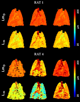 |
49 | Application of Two Denoising Methods to the Diffusion-Weighted 129Xe MRI and Dynamic Ventilation 19F/129Xe MRI Data Obtained from Normal Rats
Elise Noelle Woodward1, Gregory Lemberskiy2, Matthew S Fox1,3, and Alexei Ouriadov1,3
1Physics and Astronomy, Western University, London, ON, Canada, 2New York University Grossman School of Medicine, New York, NY, United States, 3Lawson Health Research Institute, London, ON, Canada
In this study, we demonstrate the effectiveness of two de-noising methods on diffusion-weighted and dynamic lung imaging in healthy rats using 129Xe and 19F. While the results showed a insignificant difference for denoising method 1 (MP-PCA), de-noising method 2 (MP-PCA preceded by hard-thresholding over a nuclear norm in k-space) showed significant difference in the overall mean Lm and LmD values for ventilation and r (fractional ventilation estimate) for ventilation images.
|
||
1500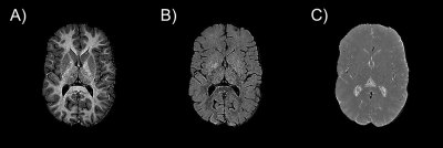 |
50 | Mapping Free Water in the Brain Using Multiparameter qMRI Combined with an Iterative Fast Exchange Relaxation Model
Ryan McNaughton1,2, Ning Hua2, Lei Zhang3, David Kennedy4, Karl Kuban2, and Hernan Jara2
1Mechanical Engineering, Boston University, Boston, MA, United States, 2Boston University Medical Center, Boston, MA, United States, 3University of North Carolina at Chapel Hill, Chapel Hill, NC, United States, 4University of Massachusetts, Worcester, MA, United States
Purpose: To develop an integrated theoretical model and computer algorithms for mapping individually and simultaneously the spatial distributions of free water, alongside myelin and iron. Theory: We solve a fast exchange relaxation model effectively decomposing qMRI maps of nPD, R1, and R2 into maps of free water, myelin, and iron. Results: The image processing technique generates maps of free water, myelin, and iron in the brains of adolescents born extremely preterm. Conclusion: A theoretical framework and image processing pipeline for mapping the distribution of free water has been developed and tested with a cohort of adolescents born extremely preterm.
|
||
1501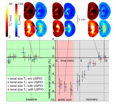 |
51 | MRI Monitoring of Changes in Kidney Size during Pathophysiological Intervention
Thomas Gladytz1, Ehsan Tasbihi1, Jason M. Millward1, Kathleen Cantow2, Luis Hummel2, Joao S. Periquito1,2, Sonia Waiczies1, Erdmann Seeliger2, and Thoralf Niendorf1,3
1Berlin Ultrahigh Field Facility (B.U.F.F.), Max Delbrueck Center for Molecular Medicine in the Helmholtz Association, Berlin, Germany, 2Institute of Physiology, Charité - Universitätsmedizin Berlin, Berlin, Germany, 3Experimental and Clinical Research Center, a joint cooperation between the Charité - Universitätsmedizin Berlin and the Max Delbrueck Center for Molecular Medicine, Berlin, Germany
Several renal pathologies are associated with changes in kidney size, offering an opportunity for magnetic resonance imaging (MRI) biomarkers of disease. An automated bean-shaped model was developed for kidney size measurements in rats using parametric MRI (T2, T2* mapping). The ABSM approach was applied to longitudinal renal size quantification during pathophysiologically relevant interventions affecting kidney size. A precision and accuracy similar to manual segmentation was achieved allowing size changes of 2% to be detected reliably. This can potentially be instrumental for developing MRI-based diagnostic tools for various kidney disorders and for gaining new insight into mechanisms of renal pathophysiology.
|
||
1502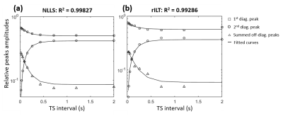 |
52 | Characterizing exchange dynamics in a urea phantom via T2-T2 relaxometry using a non-linear least squares fitting method
Ericky Caldas de A Araujo1,2, Benjamin Marty1,2, and Harmen Reyngoudt1,2
1Neuromuscular investigation center, Institute of Myology, Paris, France, 2NMR Laboratory, CEA/DRF/IBFJ/MIRCen, Paris, France
The multiexponential T2 of water in biological tissue is known to reflect microscopic anatomical compartmentation. T2-T2 correlation relaxometry allows characterizing compartmental sizes, intrinsic T2 values and exchange rates, which are of upmost clinical relevance. However, inversion of relaxation data into T2 spectra is an ill-posed problem. Regularized Inverse Laplace transform (rILT) provides stable solutions, but these are penalized by low spectral resolution and relatively high computational complexity. Here we do T2-T2 relaxometry of a urea solution and show that, for such bi-compartment system, non-linear least squares fitting provides solutions that are more accurate while avoiding the difficulties related to rILT.
|
||
1503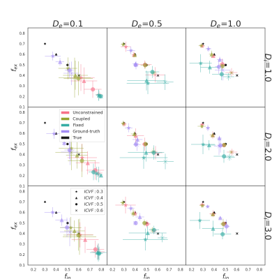 |
53 | Estimation of numerical substrate properties with compartmentalized models from Monte-Carlo simulated DW-MRI signals Video Permission Withheld
Rémy Gardier1, Juan Luis Villarreal Haro1, Erick Jorge Canales-Rodriguez1, Gabriel Girard1,2,3, Jonathan Rafael-Patiño1,3, and Jean-Philippe Thiran1,2,3
1Signal Processing Laboratory (LTS5), Ecole Polytechnique Fédérale de Lausanne, Lausanne, Switzerland, 2CIBM Center for Biomedical Imaging, Lausanne, Switzerland, 3Radiology Department, Centre Hospitalier Universitaire Vaudois and University of Lausanne, Lausanne, Switzerland
For over a decade, microstructure imaging has been a hot research topic in DW-MRI. Tissue complexity compelled researchers to make assumptions about certain properties. The inverse problem of microstructure imaging, in particular, is ill-posed, and current methods fix some parameters to reduce the solution space. In this abstract, we look at how intracellular and extracellular diffusion coefficients affect the estimation of compartmentalized model parameters. We show that robust estimation of some parameters does not extend to all parameters using Monte-Carlo simulations in impermeable substrates with multiple diffusivities, and we identified the extracellular compartment as the most influential on estimation quality.
|
||
1504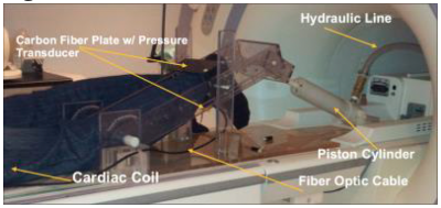 |
54 | Isometric Contractions of the Quadriceps muscle: Strain and Strain Tensor Mapping using Velocity Encoded Phase Contrast Imaging.
Shantanu Sinha1, Ryan Hernandez2, Vadim Malis1, T Oda3, and Usha Sinha2
1Radiology, UC San Diego, San Diego, CA, United States, 2Physics, San Diego State University, San Diego, CA, United States, 3Hyogo University of Teacher Education, Kato, Hyogo, Japan
The quadriceps muscle plays an extremely critical role in various aspects of human locomotion. Changes in its function from various disease such as aging, muscular disuse or modified gait can degrade mobility. VE-PC imaging provides a convenient way to study muscle motion and assess the relative contribution of the thigh muscles to force production. This study is focused on deriving the strain and strain rate tensors to explore patterns along the fiber direction and cross-section. We were able to successfully obtain strain tensor images as well as to follow the temporal variation of these indices in different muscle compartments.
|
||
1505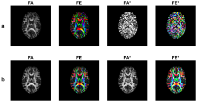 |
55 | A Hybrid IVIM-DTI Model with Optimized Acquisition Time Video Permission Withheld
Sam Sharifzadeh Javidi1 and Hamidreza Salighehrad2,3
1Department of Medical Physics and Biomedical Engineering, Tehran University of Medical Sciences, Tehran, Iran (Islamic Republic of), 2Department of Medical Physics and Biomedical Engineering, School of Medicine, Tehran University of Medical Sciences, Tehran, Iran (Islamic Republic of), 3Quantitative MR Imaging and Spectroscopy Group, Research Center for Molecular and Cellular Imaging, Tehran University of Medical Sciences, Tehran, Iran (Islamic Republic of)
IVIM-DTI imaging is capable of revealing both structural and functional maps. We conduct this study to address the long acquisition time of IVIM-DTI imaging and the inaccuracy of estimates of the model’s outputs. Diffusion-weighted images were acquired at 11 b-values and 64 orientations. We used the Kalman filter to reach higher accuracy in a lower number of images. The resulting maps indicated that diffusion maps and pseudodiffusion maps for a healthy case are of highly visual similarity. Our results also confirmed that achieving a good accuracy is possible with just half number of images.
|
||
1506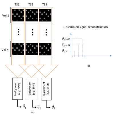 |
56 | Temporally improved volume registration by data driven subvolume interpolation of simultaneous multislice EPI time series
Oscar Albert Dabrowski1, Oscar Albert Dabrowski1, Sébastien Courvoisier2, Jean-Luc Falcone3, Antoine Klauser2, Julien Songeon3, Michel Kocher2, Bastien Chopard3, and Francois Lazeyras2
1Computer science, University of Geneva, Geneva, Switzerland, 2CIBM, Geneva, Switzerland, 3University of Geneva, Geneva, Switzerland
In the context of EPI time series acquisition, fast motion occurring below the Nyquist limit (< 1 TR), present a problem for accurate registration by 3D rigid-body algorithms. A method for temporally upsampling SMS EPI acquisitions is proposed. EPI volumes are splitted into subvolumes according to the temporal acquisition order of SMS and interpolated using a data driven approach based on pairwise affine transforms and linear interpolation. A proof of concept based EPI acquisitions of controlled head choreographies shows that fast motion below the Nyquist rate can be more accurately estimated by our method.
|
||
1507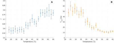 |
57 | Understanding T1 in heterogeneous systems: Extending the two-pool model to fractional order
Luke Reynolds1, Alex MacKay1,2,3, and Carl Michal1
1Physics & Astronomy, University of British Columbia, Vancouver, BC, Canada, 2Radiology, University of British Columbia, Vancouver, BC, Canada, 3UBC MRI Research Centre, University of British Columbia, Vancouver, BC, Canada
Reliably quantifying longitudinal relaxation in heterogenous systems like white matter is challenging because measured parameters are sensitive to details of the measurement technique. In particular, cross-relaxation/magnetization exchange between aqueous and non-aqueous tissue components may lead to multi-exponential relaxation during inversion recovery, depending on the difference in the pools’ initial magnetizations. We use a generalization of the Bloch equations to fractional order to show that the additional component stemming from this exchange is better described by a stretched Mittag-Leffler function than a standard exponential in heterogenous systems. This approach may provide additional information about the material structure.
|
||
1508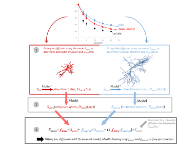 |
58 | How modeling lactate diffusion may inform on its cellular compartmentation? An initial study
Sophie Malaquin1 and Julien Valette1
1Université Paris-Saclay, Commissariat à l'Energie Atomique et aux Energies Alternatives (CEA), Centre National de la Recherche Scientifique (CNRS), Molecular Imaging Research Center (MIRCen), Laboratoire des Maladies Neurodégénératives, Fontenay aux Roses, France Brain lactate compartmentation in neurons, astrocytes and the extracellular space is presumably of critical importance, but remains impossible to assess non-invasively. DW-MRS of lactate might capture lactate fractions in each compartment, provided these compartments induce different diffusion properties. Here we propose a framework where diffusion of cell-specific metabolites (Ins and NAA) is modeled to predict lactate diffusion in astrocytes and neurons. We use some “realistic” (spheres+cylinders) and simplified (cylinders) models of cell microstructure to show that lactate fractions in each compartment can be accurately estimated, despite inaccurate microstructure modeling. Models are finally tested on in vivo mouse brain data. |
||
1509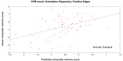 |
59 | Characterization of relationship between MR microstructural parameters and memory in older subjects
Scott Peltier1,2, Michelle Karker1, Jon-Fredrik Nielsen1,2, Navid Seraji-Bozorgzad3, Henry Paulson3, Bruno Giordani3,4, and Benjamin M. Hampstead4,5
1Biomedical Engineering, University of Michigan, Ann Arbor, MI, United States, 2Functional MRI Laboratory, University of Michigan, Ann Arbor, MI, United States, 3Neurology, University of Michigan, Ann Arbor, MI, United States, 4Psychiatry, University of Michigan, Ann Arbor, MI, United States, 5Mental Health Service, VA Ann Arbor Healthcare System, Ann Arbor, MI, United States
This work examined the relationship between white matter microstructure parameters calculated by NODDI and a composite measure of memory function. Connectome predictive modeling was done on fractional anisotropy, orientation dispersion, and neurite density maps. Significant relationships were found for all parameters, with orientation dispersion having the highest correlation between predicted and observed memory scores.
|
||
1510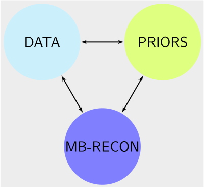 |
60 | All you need are DICOM images
Guanxiong Luo1, Moritz Blumenthal1, Xiaoqing Wang1,2, and Martin Uecker1,2,3,4
1University Medical Center Göttingen, Göttingen, Germany, 2German Centre for Cardiovascular Research (DZHK), Partner Site Göttingen, Göttingen, Germany, 3Institute of Medical Imaging, Graz University of Technology, Graz, Austria, 4Cluster of Excellence "Multiscale Bioimaging: from Molecular Machines to Networks of Excitable Cells'' (MBExC), University of Göttingen, Göttingen, Germany
Most deep-learning-based reconstructions methods need predefined sampling patterns and precomputed coil sensitivities for supervised training, limiting their later use in applications under different conditions. Furthermore, only the magnitude images are always stored in DICOM format in the Picture Archiving and Communication System (PACS) of a typical radiology department. That means that raw k-space data is usually not available. This work focuses on how to extract prior knowledge from magnitude images (DICOM) and how to apply the extracted prior to reconstruct images from k-space multi-channel data sampled with different schemes.
|
||
 Back to Meeting Home
 Back to the Program-at-a-Glance
The International Society for Magnetic Resonance in Medicine is accredited by the Accreditation Council for Continuing Medical Education to provide continuing medical education for physicians.
 Back to Meeting Home
 Back to the Program-at-a-Glance
 View the Presentation