Digital Poster
Processing & Analysis: Segmentation I
Joint Annual Meeting ISMRM-ESMRMB & ISMRT 31st Annual Meeting • 07-12 May 2022 • London, UK

Digital Poster
Processing & Analysis: Segmentation I
| Computer # | ||||
|---|---|---|---|---|
2154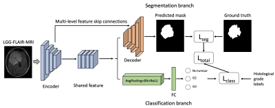 |
56 | Incorporating Histological Grading for Brain Tumor Segmentation
Lipei Zhang1, Yiran We2, Chao Li1,2, Stephen John Price2, and Carola Bibiane Schönlieb1
1The Centre of Mathematical Imaging in Healthcare, Department of Applied Mathematics and Theoretical Physics, University of Cambridge, Cambridge, United Kingdom, 2Cambridge Brain Tumor Imaging Laboratory, Division of Neurosurgery, Deoartment of Clinic Neuroscience, Univerisity of Cambridge, Cambridge, United Kingdom
Automatic tumor segmentation on MRI is crucial for patient management of brain tumors. However, previous automatic MRI segmentation methods based on deep learning are limited by bottlenecks of feature extraction. In this paper, we seek to investigate the influence of incorporating prior histology grading on the performance of tumor segmentation. We propose to employ histological grade labels to strengthen the feature extraction and tumor localization in the encoder, which can boost the DSC at exceeding 2% in different U-Net baselines. Our results could support the usefulness of incorporating clinical prior knowledge to improve the segmentation performance without introducing extra parameters.
|
||
2155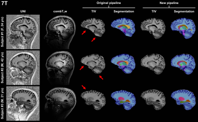 |
57 | Automated brain morphometry for sub-millimeter 7T MRI using transfer learning
Gian Franco Piredda1,2,3, Punith B. Venkategowda4, Piotr Radojewski5,6, Tom Hilbert1,2,3, Arun Joseph6,7,8, Gabriele Bonanno6,7,8, Roland Wiest5,6, Karl Egger9, Shan Yang9, Jean-Philippe Thiran2,3, Ricardo A. Corredor-Jerez1,2,3, Bénédicte Maréchal1,2,3, and Tobias Kober1,2,3
1Advanced Clinical Imaging Technology, Siemens Healthcare AG, Lausanne, Switzerland, 2Department of Radiology, Lausanne University Hospital and University of Lausanne, Lausanne, Switzerland, 3LTS5, École Polytechnique Fédérale de Lausanne (EPFL), Lausanne, Switzerland, 4Siemens Healthcare Pvt. Ltd., Bangalore, India, 5Support Center for Advanced Neuroimaging, Institute for Diagnostic and Interventional Neuroradiology, Inselspital, Bern University, Bern, Switzerland, 6Translational Imaging Center, sitem-insel AG, Bern, Switzerland, 7Advanced Clinical Imaging Technology, Siemens Healthcare AG, Bern, Switzerland, 8Magnetic Resonance Methodology, Institute of Diagnostic and Interventional Neuroradiology, University of Bern, Bern, Switzerland, 9Department of Neuroradiology, Medical Center – University of Freiburg, Faculty of Medicine, University of Freiburg, Freiburg, Germany
Large spatial signal variations due to field inhomogeneities complicate the application of automated brain morphometry at 7T. In this work, we propose to use transfer learning to adapt a template-based segmentation algorithm to sub-millimeter ultra-high field applications. More specifically, a convolutional neural network pre-trained on T1-weighted scans to extract the total intracranial volume (TIV) from MP-RAGE acquisitions was re-trained to retrieve the TIV mask directly from MP2RAGE volumes. The developed method proved to reliably deliver brain tissue masks and volumetry at 7T.
|
||
2156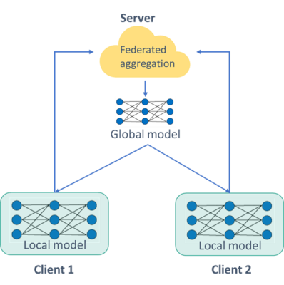 |
58 | A privacy-preserving federated learning infrastructure for prostate segmentation on T2-Weighted MRI
Fadila Zerka1, Mohammed Sunoqrot1, Bendik Abrahamsen1, Alexandros Patsanis1, Tone Frost Bathen1,2, and Mattijs Elschot1,2
1Department of Circulation and Medical Imaging, NTNU, Trondheim, Norway, 2Department of Radiology and Nuclear Medicine, St. Olavs Hospital, Trondheim, Norway
Accessing medical data is highly protected by law and ethics, making data sharing difficult and time-consuming. Distributed learning in its various forms allows learning from medical data without these data ever leaving the medical institutions. In this study, we evaluate the Flower federated learning framework for prostate segmentation on T2-Weighted MRI. The results show that the Federated learning framework performs comparably to the reference (centralized learning) model.
|
||
2157 |
59 | Reducing the impact of texture on deep-learning brain tissue segmentation networks trained with simulated MR images
Yasmina Al Khalil1, Aymen Ayaz1, Cristian Lorenz2, Jürgen Weese2, Josien Pluim1, and Marcel Breeuwer1,3
1Biomedical Engineering, Eindhoven University of Technology, Eindhoven, Netherlands, 2Philips Research Laboratories, Hamburg, Germany, 3Philips Healthcare, MR R&D - Clinical Science, Best, Netherlands
Deep learning-based segmentation algorithms largely rely on the availability of extensive, clinically representative data. While collecting such data requires vast resources, image simulation has the potential of generating realistic data reproducing a wide range of scanner sequences or parameters. In this work, we present an MR image simulation pipeline and evaluate its potential for training a deep-learning network for segmenting several brain structures in T1-weighted images acquired from real scanners. We additionally demonstrate how to prevent performance degradation from the lack of tissue texture in simulated images by combining statistical texture analysis and filtering on the evaluation image set.
|
||
2158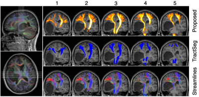 |
60 | Efficient, streamline-free white matter tract segmentation for intraoperative diffusion MRI.
Fiona Young1, Patrick Hales1,2, Laura Mancini3,4, Sotirios Bisdas3,4, Caroline Micallef3,4, Tarek Yousry3,4, Christopher A. Clark1, Kristian Aquilina5, and Jonathan D. Clayden1
1UCL Great Ormond Street Institute of Child Health, University College London, London, United Kingdom, 2Department of Radiology, Great Ormond Street Hospital for Children, London, United Kingdom, 3Lysholm Department of Neuroradiology, The National Hospital for Neurology & Neurosurgery, University College London Hospitals NHS Foundation Trust, London, United Kingdom, 4Neuroradiological Academic Unit, Department of Brain Repair and Rehabilitation, UCL Queen Square Institute of Neurology, London, United Kingdom, 5Department of Neurosurgery, Great Ormond Street Hospital for Children, London, United Kingdom
Tractfinder is an atlas-based approach to segmenting white matter tracts without tractography, in subjects with space-occupying lesions. A shape and orientation prior is combined with fibre orientation distributions in the target data to produce a map of tract location. Results for a given tract can be obtained in a matter of minutes and are comparable to those afforded by clinical tractography. In future, tractfinder could be applied to intraoperative diffusion MRI to aid neuronavigation during brain surgery.
|
||
2159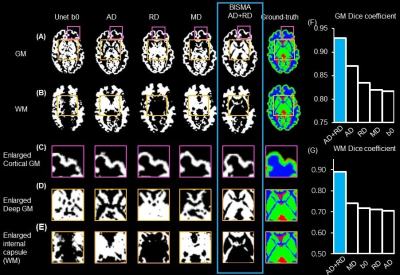 |
61 | Infant Brain Segmentation based on Multi-dMRI contrast Attention (BISMA)
Tianjia Zhu1,2, Minhui Ouyang1, Lei Feng3, Kay Sindabizera1, Jessica Hyland1, and Hao Huang1,4
1Department of Radiology, Children's Hospital of Philadelphia, Philadelphia, PA, United States, 2Department of Bioengineering, University of Pennsylvania, Philadelphia, PA, United States, 3Shandong University Cheeloo College of Medicine, Jinan, China, 4Department of Radiology, University of Pennsylvania, Philadelphia, PA, United States
On widely used relaxation-based T1-weighted (T1w) and T2-weighted (T2w) MRI images, 0-2-year-old infant brain images exhibit poor gray and white matter contrasts due to dynamic and poor myelination in this stage. Such poor T1w and T2w contrasts hinder accurate segmentation of infant brains based on T1w or T2w images. Instead, diffusion MRI (dMRI)-derived maps offer high contrasts throughout infancy. To leverage rich dMRI contrasts, we established an Infant Brain Segmentation based on Multi-dMRI contrast Attention (BISMA) technique. BISMA fills the gap in accurate deep learning segmentation of infant brain by fusing multi-contrasts of dMRI-derived maps.
|
||
2160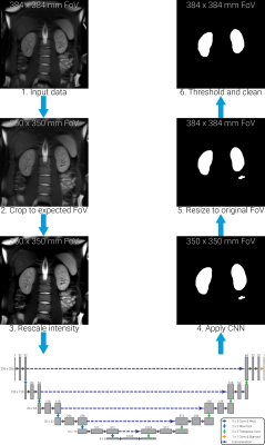 |
62 | Generalisability of Automated CNN-based Renal Segmentation for Multi-Vendor Studies
Alexander J Daniel1, Charlotte E Buchanan1, David M Morris2, Hao Li3, Rebecca Noble1, João Sousa4, Steven Sourbron4, David L Thomas5,6,7, Andrew N Priest3,8, and Susan T Francis1
1Sir Peter Mansfield Imaging Centre, University of Nottingham, Nottingham, United Kingdom, 2Centre for Cardiovascular Science, University of Edinburgh, Edinburgh, United Kingdom, 3Department of Radiology, University of Cambridge, Cambridge, United Kingdom, 4Department of Infection, Immunity and Cardiovascular Disease, University of Sheffield, Sheffield, United Kingdom, 5Neuroradiological Academic Unit, UCL Queen Square Institute of Neurology, University College London, London, United Kingdom, 6Dementia Research Centre, UCL Queen Square Institute of Neurology, University College London, London, United Kingdom, 7Wellcome Centre for Human Neuroimaging, UCL Queen Square Institute of Neurology, University College London, London, United Kingdom, 8Department of Radiology, Addenbrooke’s Hospital, Cambridge, United Kingdom
Manual segmentation of the kidneys is very time consuming and reader dependent, this renders measurements of total kidney volume (TKV) in large multi-site studies impractical. Here we use a convolutional neural network (CNN), trained on data from a single MRI vendor, to segment the kidneys of volunteers scanned with a harmonised FSE image protocol on MR scanners from three different vendors (GE, Philips and Siemens). The kidneys were manually segmented by two readers, both of which demonstrated a significant difference in TKV across vendors; no significant difference in TKV was found in the segmentations produced by the CNN.
|
||
2161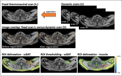 |
63 | High temporal-resolution MRI during mild-cold exposure enables the assessment of brown adipose tissue with a low inter-image variability
Aashley S.D. Sardjoe Mishre1,2, Maaike E. Straat2, Borja Martinez-Tellez2, Mariëtte R. Boon2, Oleh Dzyubachyk3,4, Andrew G. Webb1, Patrick C.N. Rensen2, and Hermien E. Kan1
1Department of Radiology, C.J. Gorter Center for High Field MRI, Leiden University Medical Center, Leiden, Netherlands, 2Department of Medicine, Division of Endocrinology and Einthoven Laboratory for Experimental Vascular Medicine, Leiden University Medical Center, Leiden, Netherlands, 3Department of Radiology, Division of Image Processing (LKEB), Leiden University Medical Center, Leiden, Netherlands, 4Department of Cell and Chemical Biology, Electron Microscopy section, Leiden University Medical Center, Leiden, Netherlands
Brown adipose tissue (BAT) is considered to be a potential therapeutic target against cardiometabolic diseases. Activated BAT combusts intracellular fatty acids leading to a reduction in fat fraction . Both cold exposure and pharmacological stimuli can activate BAT, but the short-term dynamics of BAT activation are unknown. To assess supraclavicular BAT fat fraction dynamics during cold-exposure, we developed a 1-minute-time-resolution MRI protocol using breath-holds and co-registration to minimize motion-artefacts. We demonstrated the validity and feasibility of our image analysis method, and found an inter-image variability of less than 0.1% fat fraction.
|
||
2162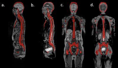 |
64 | Automatic segmentation of whole-body MRI using UnnU-Net: Feasibility of whole-skeleton ADC evaluation in plasma cell disorders
Renyang Gu1, Michela Antonelli2, Pritesh Mehta 3, Ashik Amlani 4, Adrian Green4, Radhouene Neji 5, Sebastien Ourselin2, Isabel Dregely2, and Vicky Goh2
1Department of Biomedical Engineering and Imaging Science, King's College London, London, United Kingdom, 2School of Biomedical Engineering & Imaging Sciences, King's College London, London, United Kingdom, 3University College London, London, United Kingdom, 4Department of Radiology, Guy’s & St Thomas’ NHS Foundation Trust, London, United Kingdom, 5Siemens Healthcare Limited, London, United Kingdom
Multiple myeloma is a heterogeneous bone marrow cancer. Assessment of changes in mean apparent diffusion coefficient (ADCmean) is helpful for evaluating treatment response but has not been feasible for the whole skeleton due to the time-consuming nature of manual segmentation. Whole-skeleton and per-station ADCmean were quantified from whole-body MRI using automated segmentation by an uncertainty-aware nnU-Net in 30 patients with plasma cell disorders and compared against the manual segmentation by radiologists. No differences were observed in whole-skeleton or per-station ADCmean when using the automatic and manual segmentations. Further investigation is required in a larger dataset, but initial results are promising.
|
||
2163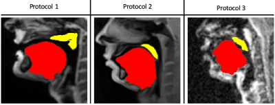 |
65 | Protocol adaptive Stacked transfer learning (STL) U-NET with small dataset training for soft tissue segmentation in dynamic speech MRI
Subin Erattakulangara1, Karthika Kelat1, and Sajan Goud Lingala1,2
1Roy J Carver Department of Biomedical Engineering, University of Iowa, Iowa city, IA, United States, 2Department of Radiology, University of Iowa, Iowa city, IA, United States
We develop a protocol adaptive Stacked transfer learning (STL) U-NET for soft tissue segmentation in dynamic speech MRI. Our approach leverages knowledge from large open-source datasets, and only needs to be trained on small number of protocol specific images (of the order of 20 images). We demonstrate the utility of STL U-NET in efficiently segmenting soft-tissue articulators from three different protocols with different field strengths, vendors, acquisition, reconstruction. Using the DICE similarity metric, we demonstrate segmentation accuracies with our approach to be at the level of manual segmentation.
|
||
2164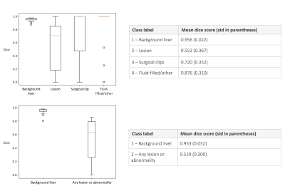 |
66 | A deep-learning non-contrast MRI lesion segmentation model for liver cancer patients that may have had previous surgery
Nora Vogt1, Zobair Arya1, Luis Núñez1, John Connell1, and Paul Aljabar1
1Perspectum, Oxford, United Kingdom Segmentation of lesions within the liver is pivotal for surveillance, diagnosis and treatment planning, and automated or semi-automated approaches can aid clinical workflows. Many patients that are under surveillance may have had previous surgery, meaning their scans will contain post-surgical features. We investigate the use of a deep learning segmentation model for non-contrast MRI that can distinguish between lesions, surgical clips and resection-induced fluid-filling regions. We report mean dice scores of 0.55, 0.72 and 0.88 for these classes respectively, demonstrating the potential of this model for semi-automated workflows. |
||
 Back to Meeting Home
 Back to the Program-at-a-Glance
The International Society for Magnetic Resonance in Medicine is accredited by the Accreditation Council for Continuing Medical Education to provide continuing medical education for physicians.
 Back to Meeting Home
 Back to the Program-at-a-Glance
 View the Presentation